User:Manpriya.A/sandbox
Introduction
[edit]Although teeth and hair are different structures in a human, they appear to develop from similar interactions between two adjacent skin layers, known as the ectoderm and mesoderm [1]. These two skin layers differentiate partly into epithelial and mesenchymal cells, respectively [1].
An essential step to understand evolution is to infer homology of different organs [2]. During tooth formation in humans, the interaction between oral epithelial cells and mesenchymal cells underneath cause 20 primary and 32 permanent teeth to form [3]. In addition, the formation of hair follicles requires skin epidermal cells, referred to as an ectodermal epithelium as well as the underlying dermal cells, known as a mesodermal mesenchyme [4]. In both the development of teeth and hair, there are interactions between epithelial and mesenchymal cells [5]. Teeth and hair share morphological similarities during very initial stages of development [5]. However, following the initial stages, similarities between tooth and hair formation generally disappear [5]. Teeth and hair share several similar developmental pathways and growth factors, including the Wnt signaling pathway, Sonic Hedgehog developmental pathway, Ectodysplasin/Ectodysplasin A Receptor pathway, and Fibroblast Growth Factors [2]. These pathways and factors are involved in forming similar structures created through epithelial-mesenchymal interactions in initial tooth and hair development [2].
Wnt Developmental Pathway
[edit]Tooth Development
[edit]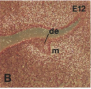
In order for the early stages of tooth development to occur through epithelial-mesenchymal interactions, the lymphoid enhancer-binding factor (Lef1) is required [6]. Lef1 is activated by the bone morphogenetic protein 4 (BMP-4) and participates in the Wnt signaling pathway [6]. The oral epithelium expresses high levels of Lef1 during the initiation of tooth formation, prior to the mesenchyme expressing Lef1 [6]. As tooth development reaches the bud stage, the mesenchyme surrounds the epithelial tooth bud invagination and subsequently begins to express Lef1 [6]. The change from epithelial cells to mesenchymal cells expressing Lef1 is consistent with the switch in the developmental dominance between these two tissues [6]. Tooth development prior to the bud stage is directed by dental epithelium, and afterwards, dental mesenchyme is the primary conductor of epithelium development towards the dental phenotype [7]. The subsequent stages of tooth development are referred to as the cap and bell stages [3]. During the cap stage, the spherical epithelial bud enlarges, gradually forming a concave surface [3]. Within the concave surface, the epithelial cells form the enamel organ region and mesenchymal cells form the dental papilla region [3]. The bell stage is characterized as the point where the epithelial cells are defined by the shape and size of the tooth they will eventually form and the dental papilla still requires Lef1 expression [3]. The presence of Lef1 transcripts in the mesenchyme and basal cells in the epithelium in both of these primary stages is required [6]. Additionally, Lef1 is expressed in pre-ameloblasts, which are derivatives of epithelial cells and are involved in the formation of enamel [6]. Therefore, the expression of Lef1 in dental epithelium and mesenchyme is necessary to prevent developmental arrest in tooth formation [6]. After the formation of the dental papilla structure, Lef1 is not required for further morphogenesis in epithelium and mesenchyme [6]. This further suggests that Lef1 expression is transient and non-cell-autonomous [6].
Hair Development
[edit]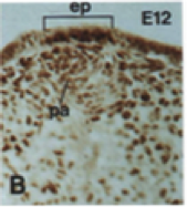
The expression of the Lef1 gene is also required in the formation of hair through epithelial and mesenchyme interactions [6]. Initially, Lef1 is detected exclusively in the mesenchyme and is used to organize the mesenchyme to encircle the epithelium [6]. Following this, additional Lef1 is expressed within the adjacent epithelium, in the basal cell layer, inducing an epithelial placode to form [6]. The individual epithelial placodes interact with the mesenchyme by inducing dermal papillae to form, which are structures involved in hair follicle nourishment [6]. After the development of dermal papillae, Lef1 is no longer necessary for further morphogenesis [6].
Similarities
[edit]
Some similarities exist between the function of Lef1 in both tooth and hair development [6]. In both teeth and hair, Lef1 is expressed in the basal cell layer of the epithelium, which is directly adjacent to the mesenchymal layer surrounding the epithelium [6]. Moreover, after the mesenchymal papilla is formed in teeth and hair, Lef1 is no longer required for further morphogenesis, despite its continuous expression [6]. Therefore, initial tooth bulb and hair follicle developmental stages have similar interactions between the mesenchymal and epithelial layers with the expression of Lef1 [6].
Sonic Hedgehog (SHH) Pathway
[edit]Tooth Development
[edit]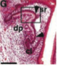
SHH is of great importance as it functions to signal the epithelial-mesenchymal interactions, which are involved in the primary stages of tooth formation [7]. During initial tooth formation, the epithelial placode of tooth germs expresses Shh [7]. In particular, incisor and molar tooth buds express Shh [8]. As development proceeds, Shh expression occurs on the tip of the budding epithelium, which is surrounded by mesenchyme during the bud stage [7]. The bud eventually forms the primary enamel knot, which is a thickened cluster of cells centralized in the inner enamel epithelium responsible for creating the crown base of the tooth [7]. Shh expression continues into the primary enamel knots, and eventually makes its way into secondary enamel knots, which are involved in the formation of cusps for teeth [7]. Furthermore, expression of Shh is transferred to ameloblasts in the bell stage [8].
Moreover, the absence of Shh during initial stages of tooth formation affects epithelial-mesenchyme interactions, resulting in small, abnormally shaped mandibular first incisors and first molars [9]. Without Shh, the maxillary incisors and molars are affected the same way, however, with lesser severity in comparison to the mandibular teeth [9]. In several instances, mandibular molars and incisors are 25% and 5% smaller, respectively [9]. Furthermore, the first molars, which usually have five cusps, sometimes only contain one cusp that is irregularly shaped, and additional cusps are affected due to a halt in their formation [9]. Additionally, the organization of the dental epithelium and mesenchyme can be abnormal due to the lack of Shh during tooth formation [9]. At the interface between epithelium and mesenchyme, the mesenchymal cells fail to form one continuous monolayer as some cells are irregularly shaped, thus impacting their ability to interact with the epithelium [9]. These results indicate that epithelial cell expression of Shh during initial stages of tooth formation is crucial for normal development [8].
Hair Development
[edit]Epithelial-mesenchymal interactions are signaled by SHH during the formation of hair follicles [7]. The development of hair includes several stages between hair germs and hair pegs, which eventually form hair follicles [7]. The hair cycle consists of three phases, known as the anagen, catagen, and telogen phases [7]. The initial anagen phase is responsible for hair follicle generation through epithelial and mesenchymal interactions [4]. During the anagen phase, the hair placode matrix epithelium cells express the Shh gene [7]. Hair germs are not capable of forming normally without expressing Shh, because without Shh expression, hair germ growth is stunted, which inhibits their elongation to form a hair peg [7]. Additionally, if Shh is not present during initial stages of hair formation, the hair bud is created in a smaller size than usual and a disorganized hair follicle results [9]. As a result, Shh is required for the proper conversion of hair germs to form into normally developed hair follicles, which involve epithelial-mesenchymal interactions [7]. Additionally, the overexpression of Shh causes an increased number of hair follicles to develop due to enhanced epithelial-mesenchymal interactions during the anagen phase [7]. Therefore, Shh is an epithelial factor, which is crucial for interactions between the epithelium and mesenchyme for hair morphogenesis [8].
Similarities
[edit]Some parallels in tooth and hair development exist including the fact that Shh expression occurs at the tip of the budding epithelium and hair placode epithelium, respectively [7]. Moreover, when Shh is not present, abnormalities in early stages of tooth and hair formation occur, preventing further normal development of teeth and hair [9].
Ectodysplasin A1 (EDA)/Ectodysplasin A Receptor (EDAR) Pathway
[edit]Mechanism of the Pathway
[edit]The Edar gene carries instructions for creating the EDAR protein, which is an essential part of a signaling pathway required for tooth formation [1]. The EDAR protein is involved in causing interactions between the ectoderm and mesoderm, which form epithelial and mesenchymal cell layers, respectively, and are both involved in creating structures such as the enamel knot and hair follicles during initial development [1]. Moreover, the Eda gene produces the protein called ectodysplasin A1, which interacts with EDAR [1]. Taken together, the ectodysplasin A1 protein interacts with the ectodysplasin A receptor, triggering signals that form structures leading to the development of tooth buds and hair follicles [1].
Tooth Development
[edit]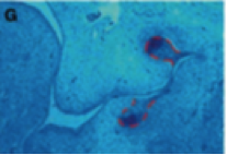
The locations where Eda and Edar are expressed vary based on the type of tooth forming within the epithelial placode, which is surrounded by mesenchyme [10]. During very early tooth development, the expression of Eda occurs after Edar expression [10]. Once Eda expression begins to increase, it is detected in the incisor tooth region located on the distal side of the epithelial thickening [10]. On the contrary, the expression of Edar occurs more proximally within the epithelial thickening [10]. The locations of Eda and Edar expression in epithelial thickenings that produce incisors is similar to that of molars, however, Eda expression is reduced in the molars [10].
During the bud stage of tooth development, Edar expression becomes increasingly restricted to the thickening dental epithelium at the bud tip, which is the region containing precursor cells for the enamel knot [10]. In the subsequent cap stage, Edar is even more strongly expressed in the enamel knot, specifically for incisors and molars, in comparison to the bud stage, and is not apparent in other epithelium regions [10]. Cusp formation, which occurs in the enamel knot, is therefore directed through this signaling pathway [11]. During the cap stage, Eda is expressed in the outer enamel epithelium at the collar region of the developing cap [10]. Therefore, the locations and patterns of expression in Edar and Eda are complementary during the cap phase of tooth formation within the epithelium [10]. Again, these expression locations are more apparent in incisor-forming regions, where Eda expression is more pronounced in epithelial cells closest to the oral cavity, and Edar is highly expressed in epithelial cells closest to the dental mesenchyme [10]. Therefore, the locations of expression of Eda and Edar in the epithelium and mesenchyme direct tooth formation [10].
Without Edar expression, the signaling pathway is disrupted, negatively affecting enamel knot formation [10]. Abnormal or missing incisors and molars are a consequence of this disruption [10]. The first molar sizes are reduced and flat compared to the normal appearance of molars [10]. Rather than deep cusps that normally form, the cusps are shallow [10]. Additionally, a reduced number of cusps in the first molars also appear in the absence of Edar [10]. Therefore, the interactions between the epithelium and mesenchyme are disrupted resulting in abnormal tooth development in the absence of Edar [10].
Hair Development
[edit]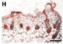
During the formation of hair follicles, Eda and Edar are both expressed in the ectoderm layer of the skin, which eventually forms epithelial cells that further, along with mesenchymal cells, produce hair follicles [12]. Further on in development when epithelial placodes begin to form, Edar expression up-regulates within the basal epithelial cells, and down-regulates in all other areas of the ectoderm [12]. These epithelial thickenings eventually form hair follicles [12]. During the period of placode formation, Eda transcripts are not apparent, although Eda is expressed in all other areas of the epidermal skin layer [12]. However, after further thickening of the epithelial bulb region of follicles occurs, Edar and Eda are co-expressed, suggesting that both are required for complete hair follicle formation [12]. Additionally, some Eda transcripts are also apparent in the mesenchyme, and along with the epithelium, help form the follicles [12].
In the absence of Eda, development of the ectoderm occurs slower than normal during the initial stages [12]. As a result, epithelial thickenings are not apparent, thus hair follicle growth is stunted [12]. These results suggest that this signaling pathway is needed for hair follicle development, which occurs through interactions between the epithelium and mesenchyme [12].
Similarities
[edit]Taken together, the EDA/EDAR signaling pathway works similarly in tooth and hair formation [12]. In both tooth and hair development, Edar is strongly expressed in the epithelial thickenings initiating the formation of the tooth bud and hair follicle [12]. Furthermore, in both scenarios, Eda expression becomes more pronounced in epithelium outside the epithelial placode where Edar expression is strongest, therefore Edar and Eda become complementary [12].
Fibroblast Growth Factor (FGF)
[edit]Tooth Development
[edit]In tooth formation, during the development of the epithelial placode, several fibroblast growth factors are expressed, suggesting that FGFs are required to initiate tooth formation [13]. The Fgf8 protein induces the expression of the Pax9 gene, which is required for tooth formation beyond the bud stage [13]. In the absence of Fgf8, there is reduced Pax9 expression in the presumptive epithelial region that forms molars, and the initiation of molar development does not occur [13]. Therefore, Fgf8 is essential for molar tooth bud development, as is the Fgf17 gene [13]. Additionally, epithelial Fgf8 controls the survival of mesenchymal cells and is a significant inductive signal within the mesenchyme for the development of molar tooth bud formation [13]. Moreover, Fgf10 is expressed in both the epithelium and mesenchyme during initial stages of tooth development, and is required for proper formation of incisor tooth buds [13]. Odontogenesis only occurs when the inducer FGF is expressed, and when the mesenchyme responds to this induction and interacts with the epithelium to initiate the bud stage of tooth formation [13]. Furthermore, the dental lamina, which is a thickening of the epithelium in areas where tooth buds develop, is formed through the epithelial expressions of Fgf8 and Fgf9 [13]. On the lingual side of the lamina, Fgf15 persists and on the tip, Fgf20 is present [13]. This indicates that Fgf8, Fgf9, Fgf15 and Fgf20 are all involved in dental lamina formation [13]. The FGFs in the dental lamina interact with the FGFs in the mesenchyme (Fgf10, Fgf18, Fgfr1lllc), causing the mesenchyme to form around the dental lamina and eventually, this leads to the formation of a tooth bud [13]. Therefore, FGF signaling is required for the interaction between epithelial and mesenchymal cells during initial stages of tooth development [13].
Hair Development
[edit]As with tooth development, the formation of structures within hair follicles requires FGFs [14]. FGFs are expressed within epithelial cells and induce mesenchymal cells to undergo mitosis [14]. Therefore, FGFs play a key role in guiding the interactions between epithelial and mesenchymal cells to form the various structures in hair follicles [14]. The different fibroblast growth factor receptors (FGFRs) and FGFs are detected in different areas of the developing hair follicle [15]. The dermal papilla within a hair follicle consists of Fgfr, which corresponds to Fgf7, while Fgfr2 is found within hair matrix epithelial cells, which extend from the hair follicle base to the tip of the hair bulb, adjacent to the dermal papilla [15]. Moreover, the hair bulb also contains Fgfr3 like Fgfr2, however, it is not detected in the base of the hair follicle [15]. Based on the locations of Fgfr3 expression, they form pre-cuticle cells [15]. Due to the lack of overlap between areas where Fgfr3 is expressed and where FGFs are detected, it is unobvious which FGFs interact with Fgfr3 [15]. The Fgfr4 gene is found in more areas of the developing hair follicle than the other three FGFRs previously described, as it is detected within the inner and outer root sheaths of the hair follicle [15]. Fgf1 binds to Fgfr4 and is involved in the differentiation of the inner root sheath [15]. All four of these FGFRs are simultaneously present during the initial anagen phase of hair formation, which involves interactions between epithelial and mesenchymal cell layers for hair follicles to develop [15]. Therefore, these ligand-receptor interactions are involved with the generation of hair follicles, which occur through epithelial-mesenchymal interactions [15].
Similarities
[edit]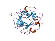
There are several different FGFs involved in tooth and hair development [13] [14] [15]. However, due to the wide variety of FGFs, different factors are expressed in different areas of the developing tooth buds and hair follicles, and are thus responsible for separate functions [13] [14] [15]. Taken together, however, both tooth and hair formation processes depend on FGFs [13] [14] [15].
Conclusion
[edit]Overall, the formation of teeth and hair occurs through interactions of two layers of the skin, the ectoderm and mesoderm [1]. These two skin layers differentiate into epithelial and mesenchymal cells, respectively, which interact with one another and secrete specific molecules to form the initial similarly formed structures in tooth and hair formation [1]. The specific molecules which are secreted from epithelial cells and/or mesenchymal cells, which are involved in both tooth and hair formation appear in several pathways and factors, including the Wnt developmental pathway, SHH pathway, EDA/EDAR signaling pathway, and FGFs [2]. Therefore, the development of tooth and hair formation is very similar as it occurs through the same developmental pathways.
References
[edit]- ^ a b c d e f g h Genetics Home Reference. (2015). EDAR. Retrieved from http://ghr.nlm.nih.gov/gene/EDAR
- ^ a b c d Sharpe, P. T. (2001). Fish scale development: hair today, teeth and scales yesterday? Current Biology, 11(18), 751-752
- ^ a b c d e Dr. Darwich, K. (n.d.). Development of teeth. Retrieved from http://iust.edu.sy/courses/7%20th%20tooth%20deve.pdf
- ^ a b Gilbert, S. F. (n.d.). Developmental Biology (10th ed.). [online]. Retrieved from http://10e.devbio.com/article.php?id=128
- ^ a b c Yildirim, S. (2013). Dental pulp stem cells. Springer Briefs in Stem Cells, 5-14
- ^ a b c d e f g h i j k l m n o p q r s Kratochwil, K., Dull, M., Farinas, I., Galceran, J., & Grosschedl, R. (1996). Lef1 expression is activated by BMP-4 and regulates inductive tissue interactions in tooth and hair development. Genes and Development, 10, 1382-1394
- ^ a b c d e f g h i j k l m n Chuong, C. M., Patel, N., Lin, J., Jung, H. S., & Widelitz, R. B. (2000). Sonic hedgehog signaling pathway in vertebrate epithelial appendage morphogenesis: perspectives in development and evolution. Cellular and Molecular Life Sciences, 57(12), 1672-1681
- ^ a b c d Iseki, S., Araga, A., Ohuchi, H., Nohno, T., Yoshioka, H., Hayashi, F., & Noji, S. (1996). Sonic hedgehog is expressed in epithelial cells during development of whisker, hair, and tooth. Biochemical and Biophysical Research Communications, 218(3), 688-693
- ^ a b c d e f g h Dassule, H. R., Lewis, P., Bei, M., Maas, R., & McMahon, A.P. (2000). Sonic hedgehog regulates growth and morphogenesis of the tooth. Development, 127, 4775-4785
- ^ a b c d e f g h i j k l m n o p q Tucker, A. S., Headon, D. J., Schneider, P., Ferguson, B. M., Overbeek, P., Tschopp, J., & Sharpe, P. T. (2000). Edar/Eda interactions regulate enamel knot formation in tooth morphogenesis. Development, 127, 4691-4700
- ^ Tucker, A. S., Headon, D. J., Courtney, J. M., Overbeek, P., & Sharpe, P. T. (2004). The activation level of the TNF family receptor, Edar, determines cusp number and tooth number during tooth development. Developmental Biology, 268(1), 185-194
- ^ a b c d e f g h i j k l Laurikkala, J., Pispa, J., Jung, H-S., Nieminen, P., Mikkola, M., Wang, X., Saarialho-Kere, U., Galceran, J., Grosschedl, R., & Thesleff, I. (2002). Regulation of hair follicle development by the TNF signal ectodysplasin and its receptor Edar. Development, 129, 2541-2553
- ^ a b c d e f g h i j k l m n o Li, C-Y., Prochazka, J., Goodwin, A. F., & Klein, O. D. (2014). Fibroblast growth factor signaling in mammalian tooth development. Odontology, 102(1), 1-13
- ^ a b c d e f Lee du Cros, D. (1993). Fibroblast growth factor and epidermal growth factor in hair development. Journal of Investigative Dermatology, 101, 106-113
- ^ a b c d e f g h i j k l Rosenquist, T. A., & Martin, G. R. (1996). Fibroblast growth factor signaling in the hair growth cycle: expression of the fibroblast growth factor receptor and ligand genes in the murine hair follicle. Developmental Dynamics, 205(4), 379-386

