User:MagdaleneLim91/sandbox
Tumefactive multiple sclerosis is a condition in which the central nervous system of a person has multiple demyelinating lesions with atypical characteristics for those of standard multiple sclerosis (MS). It is called tumefactive as the lesions are "tumor-like" and they mimic tumors clinically, radiologically and sometimes pathologically. [1]
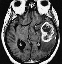
These atypical lesion characteristics include a large intracranial lesion of size greater than 2.0 cm with a mass effect, edema and a an open ring enhancement. A mass effect is the effect of a mass on the surrounding, for example, exerting pressure on the surrounding brain matter. Edema is the build-up of fluid within the brain tissue. The ring enhancement is directed toward the cortical surface. [2] The tumefactive lesion may mimic a malignant glioma or cerebral abscess causing complications in the diagnosis of tumefactive MS.


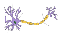
Tumefactive multiple sclerosis is a demyelinating and inflammatory disease. Myelination of the axons is highly important for signalling as it improves the speed of conduction of action potentials from one axon to the next. This is done through the formation of high-resistance, low-conductance myelin sheaths around the axons by specific cells called oligodendrocytes. As such, the demyelination process affects the communication between neurons and this consequently affects the neural pathways they control. Depending on where the demyelination takes place and how severe it is, patients with tumefactive MS have different clinical symptoms. [3]
Prevalence
[edit]Approximately 2 million people in the world suffer from multiple sclerosis and of those, more than 400,000 are from the United States[4]Tumefactive multiple sclerosis cases make up 1 to 2 of every 1000 multiple sclerosis cases. Of those cases, there are a higher percentage of females affected than males and the median age of onset is 37 years.[5]
According to the National Multiple Sclerosis Society, there are some factors that have been associated with an increased risk of MS. They include gender, ethnicity and geographic location. Based on epidemiological studies, there are about 3 times more female MS patients than male patients, indicating a possibility of an increased risk due to hormones. Among different ethnic groups, MS is the most common among Caucasians and seems to have a greater incidence at latitudes above 40° as compared to at the equator. While these associations have been made, it is still unclear how they result in an increased risk of MS onset.[6]
Genetic Factors
[edit]Studies have shown that genetic factors play a role in the increased frequency of multiple sclerosis in the relatives of affected individuals. However, identification of the genes that are involved has not been accomplished due to the small sample size.Generally, genes involved in the immune system are over-expressed and they implicate T-helper cell differentiation. T-helper cells are involved in the activation of T-cells which are responsible for entering the central nervous system and initiating demyelination processes. [7]
Clinical Signs and Symptoms
[edit]Initial symptoms of MS consist of both sensory and motor symptoms. According to Kidd, motor symptoms develop first followed by the sensory symptoms[8]The more common symptoms include spasticity, visual loss, difficulty in walking and paresthesia which is a feeling of tickling or numbness of the skin, usually described as “pins and needles”.[9]
Spasticity
[edit]Spasticity is usually the result of upper motor neuron syndrome which is caused by demyelination or inflammation in the motor areas of the brain or the spinal cord. [9] This syndrome is when motor control of skeletal muscles is affected due to damage to the efferent motor pathways. Spasticity is an involuntary muscle movement like an exaggerated stretch reflex which is when a muscle overcompensates and contracts too much in response to the muscle being stretched. It is believed that spasticity is the result of the lack of inhibitory control on the muscles, an effect of neuronal damage. [10] The upper motor neuron syndrome also includes weakness in the limbs which usually starts with the muscles at the hip and thigh thus affecting running, walking and jumping. Arm muscles usually weaken after leg muscles weaken. Some signs of weakening include loss of dexterity and stiffness of the intrinsic hand muscles.[9]
Visual Loss
[edit]Visual loss or disturbances is a result of inflammation of the optic nerve, known as optic neuritis. The effects of optic neuritis can be loss of color perception and worsening vision. Vision loss usually starts off centrally in one eye and may lead to complete loss of vision after a period of time.[9]
Cognitive Dysfunction
[edit]MS patients may show signs of cognitive impairment which is where there is a reduction in the speed of information processing, a weaker short-term memory and a difficulty in learning new concepts. [11] This cognitive impairment is associated with the loss of brain tissue, known as brain atrophy which is a result of the demyelination process in MS. [12]
Fatigue
[edit]Most MS patients experience fatigue and this could be a direct result of the disease, depression or sleep disturbances due to MS. It is not clearly understood how MS results in physical fatigue but it is known that the repetitive usage of the same neural pathways results in nerve fiber fatigue that could cause neurological symptoms. Such repeated usage of neural pathways include continuous reading which may result in temporary vision failure. [9]
Pathology
[edit]Multiple sclerosis is an autoimmune response and the breach of the blood-brain barrier by inflammatory molecules causes the demyelinating process. T lymphocytes or T-cells, involved in the immune system, are activated by binding to a specific antigen. Upon activation, the T-cells are able to penetrate through the blood-brain barrier (BBB) and enter the central nervous system (CNS). The blood-brain barrier separates blood from the cerebrospinal fluid (CSF) in the CNS. It is made of endothelial cells tightly packed together making it selectively permeable only to small molecules like carbon dioxide and oxygen. This barrier serves to protect the brain from bacterial infections. This breach of the BBB results in the increased permeability of the barrier to other inflammatory cells and macrophages. [3] Peripheral cell-signalling molecules which enter the CNS through the BBB activate microglia cells, which protect the CNS by removing infectious agents, damaged neurons and plaques. [13] Microglia cells behave like macrophages and it is believed that they are responsible for the production of the antigens that activate the T-cells and in the secretion of cytokines which may control inflammatory pathways. [14]
As of today, the initiation process of demyelination is not clearly understood but it is believed to be receptor-mediated. These macrophages attach onto the myelin sheaths and degrade the myelin, leaving the axons demyelinated. During the process of demyelination, macrophages produce toxins like nitric oxide and enzymes which cause damage to the surrounding myelin sheaths. [3] These T-cells produce high levels of tumor necrosis factor alpha (TNFα) which have been shown to be toxic towards the oligodendrocytes [13] and as such, the process of myelination is interrupted in addition to the demyelination that is occurring.
Diagnosis
[edit]Diagnosis of tumefactive MS is commonly carried out using magnetic resonance imaging (MRI), proton MR spectroscopy (H-MRS). Diagnosis is difficult as tumefactive MS may mimic the clinical and MRI characteristics of glioma or a cerebral abscess. However, as compared to tumors and abscesses, tumefactive lesions have an open-ring enhancement not a complete ring enhancement. [1] Even with this information, multiple imaging technologies have to be used together with biochemical tests for accurate diagnosis of tumefactive MS. [5]
Magnetic Resonance Imaging (MRI)
[edit]MRI diagnosis is based on lesions that are disseminated in time and space, meaning that there are multiple episodes and consisting of more than one area. [15] There are two kinds of MRI used in the diagnosis of tumefactive MS, T1-weighted imaging and T2-weighted imaging. Using T1-weighted imaging, the lesions are displayed with low signal intensity, meaning that the lesions appear darker than the rest of the brain. Using T2-weighted imaging, the lesions appear with high signal intensity, meaning that the lesions appear white and brighter than the rest of the brain. When T1-weighted imaging is contrast-enhanced through the addition of gadolinium, the open ring enhancement can be viewed as a white ring around the lesion. [16]
Proton MR Spectroscopy (H-MRS)
[edit]H-MRS identifies biochemical changes in the brain such as the quantity of metabolic products of neural tissue including choline, creatine, N-acetylaspartate (NAA), mobile lipids and lactic acid. When demyelination is occurring, there is breakdown of cell membranes resulting in an increase in the level of choline. NAA is specific to neurons and thus, a reduction in NAA concentration indicates neuronal or axonal dysfunction. As such, the levels of choline and NAA can be measured to determine if there is demyelination activity and inflammation in the brain. Usually, the ratio of choline to NAA is used. [17]
Treatment
[edit]Pharmacologic
[edit]Disease-modifying Agents
[edit]Pharmacologic treatments for MS include immunomodulators and immunosuppresants which reduce the frequency and severity of relapses by about 35% and reduce the lesion growth. [18]
Interferon beta (IFN-beta)
[edit]IFN-beta one of the first treatments available for MS and is an immunomodulator as it alters antigen presentation and affects the proliferation of T cells. As explained in the pathology of MS, the binding of an antigen to the T-cell results in its activation and passes through the BBB, allowing inflammatory cells and macrophages to enter the CNS and initiate demyelination. As such, IFN-beta has proven to decrease the rate of relapses.
The presence of neutralizing antibodies against IFN-beta in the body will oppose the activity of IFN-beta and thus, IFN-beta treatment will have no effect in certain individuals who produce those neutralizing antigens. There are many known side-effects of IFN-beta. They include flu-like symptoms, leukopenia and an increased levels of liver enzyme. [19]
Glatiramer acetate has proven to affect the differentiation of T-cells as well as altering the activity of antigen presenting cells and restoring the deficiency of certain proteins like CD4 in T-cells. These T-cells then become less able to get activated by an antigen, preventing a relapse in MS from happening. Currently, no side effects of this treatment have been found. [19]
Mitoxantrone is an immunosuppressant drug and prevents the proliferation of T-cells by inhibiting DNA repair proteins and DNA topoisomerase which are vital in the DNA replication process before mitosis. However, mitoxantrone can cause damage to the heart and as such, it is usually administered to MS patients whose disease is at a rapid progressive rate. [20]
Corticosteroids have immunosuppressive and anti-inflammatory properties and are used in MS therapy as they restore the blood-brain barrier and induce cell death of T-cells.[20]
Dietary Supplementaion
[edit]Vitamin D is believed to affect the growth and differentiation of macrophages and T cells and as such, is used as treatment for MS. Higher levels of vitamind D have also proven to reduce the risk of the onset of MS. [2]
Treatment of Symptoms
[edit]Due to the wide range of symptoms experienced by patients with MS, the treatment for each MS patient varies depending on the extent of the symptoms.
Treatment of Spasticity
[edit]The treatment of spasticity ranges from physical activity to medication. Physical activity includes stretching, aerobic exercises and relaxation techniques. Currently, there is little understanding as to why these physical activities aid in relieving spasticity. Medical treatments include baclofen, diazepam and dantrolene which is a muscle-relaxant. Dantrolene has many side effects and as such, it is usually not the first choice in treatment of spasticity. The side effects include dizziness, nausea and weakness. [20]
Treatment of Fatigue
[edit]Fatigue is a common symptom and affects the daily life of individuals with MS. Changes in lifestyle are usually recommended to reduce fatigue such as taking frequent naps and implementation of exercise. MS patients who smoke are advised to stop. Pharmacological treatment include anti-depressants and caffeine. Aspirin has also been experimented with and from clinical trial data, MS patients preferred using aspirin as compared to the placebo in the test. One hypothesis is that aspirin has an effect on the hypothalamus and can affect the perception of fatigue through altering the release of neurotransmitters and the autonomic responses. [20]
Treatment of Cognitive Dysfunction
[edit]There are no approved drugs for the treatment of cognitive dysfunction, however, some treatments have shown an association with improvements in cognitive function. One such treatment is Ginkgo biloba, which is a herb commonly used by patients with Alzheimer's disease.[20]
See also
[edit]References
[edit]- ^ a b Xia, L., Lin, S., Wang, Z., Li, S., Xu, L., Wu, J., Hao, S. and Gao, C. Tumefactive demyelinating lesions: nine cases and a review of the literature. Neurosurg Rev 32:171-179 (2009).
- ^ a b Kaeser, M. A., Scali, F., Lanzisera, F. P., Bub, G. A., and Kettner, N. W. Tumefactive multiple sclerosis: an uncommon diagnostic challenge. Journal of Chiropractic Medicine 10:29-35 (2011).
- ^ a b c Moore, G. R. W., and Esiri, M. M. The pathology of multiple sclerosis and related disorders. Diagnostic Histopathology 17(5):225-231.
- ^ Peterson, J. W., Trapp, B. D. Neuropathobiology of multiple sclerosis. Neurol Clin, 2005; 23(1):107–129
- ^ a b Lucchinetti, C. F., Gavrilova, R. H., Metz, I., Parisi, J. E., Scheithauer, B. W., Weigand, S., et al. Clinical and radiographic spectrum of pathologically confirmed tumefactive multiple sclerosis. Brain 131(7):1759-1775 (2008).
- ^ National Multiple Sclerosis Society. "Epidemiology of MS". Retrieved 11/17/2012.
{{cite web}}: Check date values in:|accessdate=(help) - ^ Dyment, D. A., Yee, I. M., Ebers, G. C., and Sadovnick, A. D. Multiple sclerosis in stepsiblings: recurrence risk and ascertainment. J. Neurol. Neurosurg. Psychiatry 77:258-259 (2006)
- ^ Kidd PM. Multiple sclerosis, an autoimmune inflammatory disease: prospects for its integrative management. Altern Med Rev 2001; 6(6):540-66.
- ^ a b c d e Rudick, R. A. Contemporary Diagnosis and Management of Multiple Sclerosis. Pennsylvania: Handbooks in Health Care Co., 2004. Print.
- ^ Purves, D., Augustine, G. J., Fitzpatrick, D., Hall, W. C., LaMantia, A. S., and White, L. E. Neuroscience. Sunderland: Sinauer Associates Inc.,U.S., 2010. Print.
- ^ Crayton, H. J., and Rossman, H. S. Managing the Symptoms of Multiple Sclerosis: A Multimodal Approach. Clinical Therapeutics 28(4): 445-460(2006)
- ^ Pelletier, J., , Suchet L., and Witjas T., et al. A longitudinal study of callosal atrophy and interhemispheric dysfunction in relapsing-remitting multiple sclerosis. Arch Neurol 58: 105-111 (2001).
- ^ a b Lindquist, S., Hassinger, S., Lindquist, J. A., and Sailer, M. The balance of pro-inflammatory and trophic factors in multiple sclerosis patients: effects of acute relapse and immunomodulatory treatment. Multiple Sclerosis Journal 17(7):851-866.
- ^ Deng, X., and Sriram, S. Role of Microglia in Multiple Sclerosis. Current Neurology and Neuroscience Reports 5:239-244 (2005).
- ^ Tintore, M., Rovira, A., Martinex, M. J., Rio, J., diaz-Villoslada, P., Brieva, L., et al. Isolated demyelinating syndromes: comparison of different MR imaging criteria to predict conversion to clinically definite multiple sclerosis. AJNR AM J Neuroradiol; 21(4):702-706 (2000).
- ^ Takeuchi, T., Ogura, M., Sato, M., Kawai, N., Tanihata, H., Takasaka, I., Minamiguchi, H., Nakai, M., and Itakura, T. Late-onset tumefactive multiple sclerosis. Radiat Med 26:549-552 (2008).
- ^ Sajja, B. R., Wolinsky, J. S., Narayana, P. A. Proton magnetic resonance spectroscopy in multiple sclerosis. Neuroimaging Clin N Am; 19(1):45-58 (2009).
- ^ White LJ, Dressendorfer RH. Exercise and multiple sclerosis. Sports Med; 34(15):1077-100 (2004).
- ^ a b Buck, D. and Hemmer, B. Treatment of multiple sclerosis: current concepts and future perspectives. J Neurol 258: 1747-1762 (2011).
- ^ a b c d e Crayton, H. J., and Rossman, H. S. Managing the Symptoms of Multiple Sclerosis: A Multimodal Approach. Clinical Therapeutics 28(4) (2006).
