User:CookingandMedicine/Ludwig's angina
| This is the sandbox page where you will draft your initial Wikipedia contribution.
If you're starting a new article, you can develop it here until it's ready to go live. If you're working on improvements to an existing article, copy only one section at a time of the article to this sandbox to work on, and be sure to use an edit summary linking to the article you copied from. Do not copy over the entire article. You can find additional instructions here. Remember to save your work regularly using the "Publish page" button. (It just means 'save'; it will still be in the sandbox.) You can add bold formatting to your additions to differentiate them from existing content. |
Treatment
[edit]For each patient, the treatment plan should be consider the patient's stage of infection, airway control, and comorbidities. Other things to consider include physician experience, available resources, and personnel are critical factors in formulation of a treatment plan.[1] There are four principles that guide the treatment of Ludwig's Angina:[2] Sufficient airway management, early and aggressive antibiotic therapy, incision and drainage for any who fail medical management or form localized abscesses, and adequate nutrition and hydration support. Each will be explained in detail below.[citation needed]
Airway management
[edit]
Airway management has been found to be the most important factor in treating patients with Ludwig's Angina,[3] i.e. it is the “primary therapeutic concern”.[4] Airway compromise is known to be the leading cause of death from Ludwig's Angina.[5]
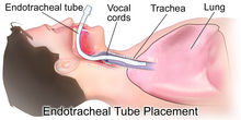
- The basic method to achieve this is to allow the patient to sit in an upright position with supplemental oxygen provided by masks or nasal prongs.[3] Patient's airway can rapidly deteriorate and therefor close observation and preparation for more invasive methods such as endotracheal intubation or tracheostomy[3] if needed is vital.
- If the oxygen saturation levels are adequate and antimicrobials have been given, simple airway observation can be done.[3] This is a suitable method to adopt in the management of children, as a retrospective study described that only 10% of children required airway control. However, a tracheostomy was performed on 52% of those affected with Ludwig's Angina over 15 years old.[6]
- If more invasive or surgical airway control is necessary, there are multiple things to consider[5]
- Flexible nasotracheal intubation require skills and experience.[5]
- If nasotracheal intubation is not possible, cricothyrotomy and tracheostomy under local anaesthetic can be done. This procedure is carried out on patients with advanced stage of Ludwig's Angina.[5]
- Endotracheal intubation has been found to be in association with high failure rate with acute deterioration in respiratory status.[5]
- Elective tracheostomy is described as a safer and more logical method of airway management in patients with fully developed Ludwig's Angina.[7]
- Fibre-optic nasoendoscopy can also be used, especially for patients with floor of mouth swellings.[3]
Antibiotics
[edit]- Antibiotic therapy is empirical, it is given until culture and sensitivity results are obtained.[3] The empirical therapy should be effective against both aerobic and anaerobic bacteria species commonly involved in Ludwig's Angina.[3] Only when culture and sensitivity results return should therapy be tailored to the specific requirements of the patient.[3]
- Empirical coverage should consist of either a penicillin with a B-lactamase inhibitor such as amoxicillin/ticarcillin with clavulanic acid or a Beta-lactamase resistant antibiotic such as cefoxitin, cefuroxime, imipenem or meropenem.[3] This should be given in combination with a drug effective against anaerobes such as clindamycin or metronidazole.[3]
- Parenteral antibiotics are suggested until the patient is no longer febrile for at least 48 hours.[3] Oral therapy can then commence to last for 2 weeks, with amoxicillin with clavulanic acid, clindamycin, ciprofloxacin, trimethoprim-sulfamethoxazole, or metronidazole.[3]
Incision and drainage
[edit]- Surgical incision and drainage are the main methods in managing severe and complicated deep neck infections that fail to respond to medical management within 48 hours.[3]
- It is indicated in cases of:[3]
- Airway compromise
- Septicaemia
- Deteriorating condition
- Descending infection
- Diabetes mellitus
- Palpable or radiographic evidence of abscess formation
- Bilateral submandibular incisions should be carried out in addition to a midline submental incision. Access to the supramylohyoid spaces can be gained by blunt dissection through the mylohyoid muscle from below.[3]
- Penrose drains are recommended in both supramylohyoid and inframylohyoid spaces bilaterally. In addition, through and through drains from the submandibular space to the submental space on both sides should be placed as well.[3]
- The incision and drainage process is completed with the debridement of necrotic tissue and thorough irrigation.[3]
- It is necessary to mark drains in order to identify their location. They should be sutured with loops as well so it will be possible to advance them without re-anaesthetizing the patient while drains are re-sutured to the skin.[3]
- An absorbent dressing is then applied. A bandnet dressing retainer can be constructed so as to prevent the use of tape.[3]
Other things to consider:
[edit]Nutritional support
[edit]Adequate nutrition and hydration support is essential in any patient following surgery, particularly young children.[2] In this case, pain and swelling in the neck region would usually cause difficulties in eating or swallowing, hence reducing patient's food and fluid intake. Patients must therefore be well-nourished and hydrated to promote wound healing and to fight off infection.[8]
Post-operative care
[edit]Extubation, which is the removal of endotracheal tube to liberate the patient from mechanical ventilation, should only be done when the patient's airway is proved to be patent, allowing adequate breathing. This is indicated by a decrease in swelling and patient's capability of breathing adequately around an uncuffed endotracheal tube with the lumen blocked.[8]
During the hospital stay, patient's condition will be closely monitored by:
- carrying out cultures and sensitivity tests to decide if any changes need to be made to patient's antibiotic course
- observing for signs of further infection or sepsis including fevers, hypotension, and tachycardia
- monitoring patient's white blood cell count - a decrease implies effective and sufficient drainage
- repeating CT scans to prove patient's restored health status or if infection extends, the anatomical areas that are affected.[8]
Moreover, it is advised to never leave young children with significant neck swelling unattended and they should always be seated to prevent suffocation.[2]
References
[edit]- ^ Shockley WW (May 1999). "Ludwig angina: a review of current airway management". Archives of Otolaryngology–Head & Neck Surgery. 125 (5): 600. doi:10.1001/archotol.125.5.600. PMID 10326825.
- ^ a b c Chou YK, Lee CY, Chao HH (December 2007). "An upper airway obstruction emergency: Ludwig angina". Pediatric Emergency Care. 23 (12): 892–6. doi:10.1097/pec.0b013e31815c9d4a. PMID 18091599. S2CID 2891390.
- ^ a b c d e f g h i j k l m n o p q r s Bagheri SC, Bell RB, Khan HA (2011). Current Therapy in Oral and Maxillofacial Surgery. Philadelphia: Elsevier. pp. 1092–1098. ISBN 978-1-4160-2527-6.
- ^ Moreland LW, Corey J, McKenzie R (February 1988). "Ludwig's angina. Report of a case and review of the literature". Archives of Internal Medicine. 148 (2): 461–6. doi:10.1001/archinte.1988.00380020205027. PMID 3277567.
- ^ a b c d e Saifeldeen K, Evans R (March 2004). "Ludwig's angina". Emergency Medicine Journal. 21 (2): 242–3. doi:10.1136/emj.2003.012336. PMC 1726306. PMID 14988363.
- ^ Kurien M, Mathew J, Job A, Zachariah N (June 1997). "Ludwig's angina". Clinical Otolaryngology and Allied Sciences. 22 (3): 263–5. doi:10.1046/j.1365-2273.1997.00014.x. PMID 9222634.
- ^ Parhiscar A, Har-El G (November 2001). "Deep neck abscess: a retrospective review of 210 cases". The Annals of Otology, Rhinology, and Laryngology. 110 (11): 1051–4. doi:10.1177/000348940111001111. PMID 11713917. S2CID 40027551.
- ^ a b c Bagheri SC, Bell RB, Khan HA (2012). Current Therapy in Oral and Maxillofacial Surgery. Philadelphia: Elsevier Saunders. ISBN 978-1-4160-2527-6. OCLC 757994410.
