Mandibular setback surgery
Mandibular setback surgery is a surgical procedure performed along the occlusal plane to prevent bite opening on the anterior or posterior teeth and retract the lower jaw for both functional and aesthetic effects in patients with mandibular prognathism.[1][2] It is an orthodontic surgery that is a form of reconstructive plastic surgery.[3] There are three main types of procedures for mandibular setback surgery: Bilateral Sagittal Split Osteotomy (BSSO), Intraoral Vertical Ramus Osteotomy (IVRO) and Extraoral Ramus Osteotomy (EVRO), depending on the magnitude of mandibular setback for each patient. Postoperative care aims to minimise postoperative complications, complications includes bite changes, relapse and nerve injury.

History
[edit]The origin of orthognathic surgery was introduced by Simon P. Hullihen in 1849.[4] Upon Hullihen's discovery of the procedure, many surgeons further investigated and modified approaches to correct mandibular prognathism.[5] Vilray Blair and Edward H. Angle collaborated to introduce osteotomy to the mandibular body with a horizontal incision, marking the beginning of orthognathic surgery.[6] With these notable modifications in surgical procedures for prognathism, BSSO, IVRO and EVRO are procedures used in practise to treat individuals with dentofacial deformities. VRO was first introduced as EVRO by Caldwell and Letterman and further modified into IVRO by Moose in 1964.[7][8][9][10] BSSO was then introduced by Obwegeser in 1959.[11]
Anatomy
[edit]
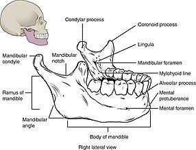
Mandible is a movable solitary bone forming the lower jaw.[12] It is composed of a mandibular body and two mandibular rami (singular: ramus[13]) on bilateral sides of the face.[14] The mandibular ramus contains the mandibular foramen, which is where the inferior alveolar nerve and artery enters.[14] Each end of the ramus has a projection that articulates with the temporal bones via temporomandibular joint called mandibular condyle.[14]
Lingula is superior to mandibular foramen on the mandibular ramus, which often acts as a recognisable feature for surgical procedures.[15][16]
Surgical procedures
[edit]BSSO and IVRO are the two most common types of procedures used for mandibular setback surgery while EVRO is carried out for specific cases.
Bilateral Sagittal Split Osteotomy (BSSO)
[edit]
A vertical incision is performed on the inferior and lateral sides of the soft tissue in the mouth at a distance from the adjacent gums.[17] The cut is performed from the mandibular ramus to the mandibular body along the external oblique line, down to the mandibular first molar region, and further down the buccal vestibule of the mandible.[16] A precise cut allows sufficient tissues remained for closing the suture.[17] The surrounding soft tissue from the bone is dissected and elevated in a subperiosteal plane to see the mandibular ramus clearly. The mandibular foramen and the lingula must also be exposed without damaging the inferior alveolar nerve. A clamp is used to hold the layers in a fixed position for mandibular osteotomy to be carried out.

The BSSO technique requires two cuts of the mandible utilising an osteotome that is inclined to one side.[17] Firstly, the lateral osteotomy starts at the buccal cortex, the bone in the buccal space. This split is done vertically down to the first or second molar region.[18] Then the medial osteotomy is done on the lingual cortical bone, which extends to the anterior border of the ramus posterior and to the inferior alveolar canal.[16] Subsequently, the fractures of the ramus are split by applying slight force.
Mobilisation of the segments are done to the pre-arranged position and fixed internally with screws and plates.[19] BSSO allows rigid fixation and increases stability of mandibular setbacks. However, as the nerve is manipulated to a large extent during the surgery, it increases the chance of patients experiencing neurosensory loss.[20] Studies have shown statistically that another type of method, IVRO, can reduce the chance of neurosensory disturbance.[21]
Intraoral Vertical Ramus Osteotomy (IVRO)
[edit]
Firstly, a horizontal incision through the oral mucosa along the anterior border of the ramus of mandible within the mouth allows surgeons to reach the underlying bone and tissue of the lower jaw.[16] Once the cut opens near the coronoid process and mandibular first molar region, a deeper dissection is done so the mandibular notch, inferior and posterior of the ramus are visible.[16] Then, the IVRO can be performed.
A pair of retractors are positioned properly and carefully for clear exposure of the ramus.[16] The cutting of the bone is done by surgical oscillating saw,[22] at an angle of 105° precisely behind the body prominence of antilingula, superior to the sigmoid notch and inferior to the mandibular angle. The bone fragment is adjusted to the preoperative planned position, which sets the lower jaw backwards.[23] Stabilisation of the jaw at its modified position is then done through wiring. The inter-maxillary fixation is usually kept for around six weeks.[16]
The IVRO procedure can be performed if the magnitude of the mandibular setback is within 10mm.[24] The limitation of the specific range of setback is to prevent postoperative forward relapse and the dislocation of mandibular condylar head.[24] When the required adjustment of the jaw exceeds 10mm, patients will most likely undertake the EVRO procedure.
Extraoral Vertical Ramus Osteotomy (EVRO)
[edit]The incision for EVRO is made on the most superficial layer of the skin, approximately 1 to 2 cm below the mandibular angle.[25][26] The dissection cuts the platysma and the masseter muscle. As the skin dissection is done, a small protrude of the ramus could be observed. This allows the mandible osteotomy to be done safely by avoiding damaging the adjacent nerves.[16]
The bony cut is made from the sigmoid notch to the mandibular angle with an osteotome. The bony fragment at the proximal end is rearranged and stabilized using screws, plates, or wires. The closure of the wound is done layer by layer. The intermaxillary fixation is usually kept for a week to ten days.[25]
Compared to BSSO and IVRO, this technique takes the least surgical time as it is performed on the external surface, which allows the surgeons to have easier access and clearer vision. However, the surgical scar that is left behind could be a concern to certain patients.
Effects
[edit]The mandibular setback surgery can improve masticatory function and facial aesthetics by repositioning the jaw and mouth, which can have a positive effect on the patient's psychosocial well-being.[27][28]
Functional
[edit]Patients with mandibular prognathism may have difficulty chewing, speaking, and breathing, which can affect their quality of life.[17] The mandibular setback surgery improves one's masticatory muscle activity and speech intelligibility.[29] The mandibular setback surgery improves functional difficulties caused by mandibular prognathism.
Aesthetic
[edit]Aesthetic correction is second positive effect of mandibular setback surgery. In particular, it improves the asymmetry of the upper and lower lip in patients with mandibular prognathism. Patients after the surgery may result in a more pronounced and defined chin by repositioning the jaw.[30][31] Ultimately, it allows for a more balanced and harmonious facial profile.[32]
Psychological
[edit]Post-surgery questionnaires results indicated improved psychosocial well-being, self-esteem and social functioning in patients after the mandibular setback surgery.[32] There is also a high patient satisfaction rate for the surgery.[33] These show how the mandibular setback surgery improves well-being of patients with mandibular prognathism.[28]
Post-operative care
[edit]After the setback surgery, a 2-week recovery period is needed for the wires to be closed and inter-maxillary fixation to stabilise the position of the mandible.[34] Patients would stay for a minimum of 1 to 2 days and exception may occur if a complication has occurred. Post-operative assessments need to be done for the 2 weeks after the operation.
Gum chewing and patient education exercise post-surgery can improve masticatory function in patient and minimise the risk of complications.[35][36]
Risk and complications
[edit]More than 40% of patients have complications after mandibular setback surgery.[31] Complications include bite changes, nerve injuries and relapse.
Bite changes
[edit]Bite changes occur in 20.3% of the cases post-setback surgery.[37] Change in pharyngeal airway space and tongue position can have a significant effect on bite changes after mandibular setback surgery and cause obstructive sleep apnea.[1][38][39]
The tongue is normally positioned against the roof of the mouth, supporting the upper jaw. After surgery, the change in the position of the tongue affects the position of the jaw, leading to bite changes.[40] Bite changes can narrow the pharyngeal airway space after surgery can lead obstructive sleep apnea.[41] Incorrect jaw positioning requires additional surgery to reposition the jaw and open up the airway.[42]
Relapse
[edit]9.2% of the patients who underwent the mandibular setback surgery has found post-surgery relapse.[43] This is caused by actions of the condyle resorption.[44] Condyle resorption is when the bone tissue is lost in the condyle. Condyle resorption reduces stability of the mandible and cause long term skeletal relapse.[45][46] When the mandible is not stabilised, it allows movement of the mandible into its pre-operative position, contributing to early relapse.[47] Bone fixation procedures will be needed due to the lack of bone healing.[48]
Nerve injuries
[edit]Nerve injuries occur in 3.7% of the patients after the mandibular setback surgery.[49] Cutting and repositioning of the mandible in the surgery can potentially damage nerves in the mandible that is responsible for sensation and movement. Specifically, the inferior alveolar nerve are the commonly affected nerve in the surgery.[50] Less common nerves injuries are on the lingual nerve and mental nerve, which are responsible for tongue and chin sensation respectively. The lingual nerve is affected by the wire placement in the molar region.[51] The mental nerve injury can be caused by the presence of bony spurs. A damage in the nerve may require additional therapy to repair the loss of ability in the nerves.[52]
References
[edit]- ^ a b Lee K, Hwang SJ (December 2019). "Change of the upper airway after mandibular setback surgery in patients with mandibular prognathism and anterior open bite". Maxillofacial Plastic and Reconstructive Surgery. 41 (1): 51. doi:10.1186/s40902-019-0230-4. PMC 6877677. PMID 31824889.
- ^ Proffit WR, Phillips C, Dann C, Turvey TA (1991). "Stability after surgical-orthodontic correction of skeletal Class III malocclusion. I. Mandibular setback". The International Journal of Adult Orthodontics and Orthognathic Surgery. 6 (1): 7–18. PMID 1940541.
- ^ "Is orthognathic surgery plastic surgery?". www.aestheticjawsurgery.com. Retrieved 2023-04-12.
- ^ Kashani H, Rasmusson L (2016-08-31). "Osteotomies in Orthognathic Surgery". In Motamedi MH (ed.). A Textbook of Advanced Oral and Maxillofacial Surgery. Vol. 3. InTech. doi:10.5772/63345. ISBN 978-953-51-2590-7. S2CID 4677420. Retrieved 2023-04-13.
- ^ Aziz SR (October 2004). "Simon P. Hullihen and the origin of orthognathic surgery". Journal of Oral and Maxillofacial Surgery. 62 (10): 1303–1307. doi:10.1016/j.joms.2003.08.044. PMID 15452820.
- ^ Böckmann R, Meyns J, Dik E, Kessler P (December 2014). "The modifications of the sagittal ramus split osteotomy: a literature review". Plastic and Reconstructive Surgery. Global Open. 2 (12): e271. doi:10.1097/GOX.0000000000000127. PMC 4292253. PMID 25587505.
- ^ Chen CM, Lee HE, Yang CF, Shen YS, Huang IY, Tseng YC, Lai ST (July 2008). "Intraoral vertical ramus osteotomy for correction of mandibular prognathism: long-term stability". Annals of Plastic Surgery. 61 (1): 52–55. doi:10.1097/sap.0b013e318153f3ee. PMID 18580150. S2CID 25472775.
- ^ Ayoub AF, Millett DT, Hasan S (August 2000). "Evaluation of skeletal stability following surgical correction of mandibular prognathism". The British Journal of Oral & Maxillofacial Surgery. 38 (4): 305–311. doi:10.1054/bjom.2000.0303. PMID 10922156.
- ^ Jung HD, Jung YS, Park HS (April 2009). "The chronologic prevalence of temporomandibular joint disorders associated with bilateral intraoral vertical ramus osteotomy". Journal of Oral and Maxillofacial Surgery. 67 (4): 797–803. doi:10.1016/j.joms.2008.11.003. PMID 19304037.
- ^ Jacobovicz J, Lee C, Trabulsy PP (February 1998). "Endoscopic repair of mandibular subcondylar fractures". Plastic and Reconstructive Surgery. 101 (2): 437–441. doi:10.1097/00006534-199802000-00030. PMID 9462779.
- ^ Troulis MJ, Kaban LB (July 2004). "Endoscopic vertical ramus osteotomy: early clinical results". Journal of Oral and Maxillofacial Surgery. 62 (7): 824–828. doi:10.1016/j.joms.2003.12.021. PMID 15218560.
- ^ Singh, Gagandeep. "Solitary bone cyst of the mandible | Radiology Reference Article | Radiopaedia.org". Radiopaedia.
- ^ "Ramus of mandible - e-Anatomy - IMAIOS".
- ^ a b c Breeland G, Aktar A, Patel BC (2023). "Anatomy, Head and Neck, Mandible". StatPearls. Treasure Island (FL): StatPearls Publishing. PMID 30335325. Retrieved 2023-04-13.
- ^ Lupi SM, Landini J, Olivieri G, Todaro C, Scribante A, Rodriguez Y, Baena R (December 2021). "Correlation between the Mandibular Lingula Position and Some Anatomical Landmarks in Cone Beam CT". Healthcare. 9 (12): 1747. doi:10.3390/healthcare9121747. PMC 8701814. PMID 34946470.
- ^ a b c d e f g h Mani V (2021). "Orthognathic Surgery for Mandible". In Bonanthaya K, Panneerselvam E, Manuel S, Kumar VV (eds.). Oral and Maxillofacial Surgery for the Clinician. Singapore: Springer Nature. pp. 1477–1512. doi:10.1007/978-981-15-1346-6_68. ISBN 978-981-15-1346-6. S2CID 234341270.
- ^ a b c d Prasad V, Kumar S, Pradhan H, Siddiqui R, Ali I (2021). "Bilateral sagittal split osteotomy a versatile approach for correction of facial deformity: A review literature". National Journal of Maxillofacial Surgery. 12 (1): 8–12. doi:10.4103/njms.NJMS_89_18. PMC 8191559. PMID 34188394.
- ^ profilbaru.com. "Orthognathic surgery - Profilbaru.Com". Retrieved 2023-04-13.
- ^ Themes UF (2017-01-23). "The Bilateral Sagittal Split Mandibular Ramus Osteotomy". Pocket Dentistry. Retrieved 2023-04-13.
- ^ Roychoudhury S, Nagori SA, Roychoudhury A (May 2015). "Neurosensory disturbance after bilateral sagittal split osteotomy: A retrospective study". Journal of Oral Biology and Craniofacial Research. 5 (2): 65–68. doi:10.1016/j.jobcr.2015.04.006. PMC 4523587. PMID 26258016.
- ^ Al-Moraissi EA, Ellis E (July 2015). "Is There a Difference in Stability or Neurosensory Function Between Bilateral Sagittal Split Ramus Osteotomy and Intraoral Vertical Ramus Osteotomy for Mandibular Setback?". Journal of Oral and Maxillofacial Surgery. 73 (7): 1360–1371. doi:10.1016/j.joms.2015.01.010. PMID 25871900.
- ^ "What is an surgical oscillating saw?".
- ^ Rokutanda S, Yamada SI, Yanamoto S, Sakamoto H, Furukawa K, Rokutanda H, et al. (March 2020). "Anterior relapse or posterior drift after intraoral vertical ramus osteotomy". Scientific Reports. 10 (1): 3858. Bibcode:2020NatSR..10.3858R. doi:10.1038/s41598-020-60838-1. PMC 7052185. PMID 32123263.
- ^ a b Themes UF (2016-06-03). "Intraoral Vertical Ramus Osteotomy". Pocket Dentistry. Retrieved 2023-04-13.
- ^ a b Öhrnell Malekzadeh B, Ivanoff CJ, Westerlund A, MadBeigi R, Öhrnell LO, Widmark G (February 2022). "Extraoral vertical ramus osteotomy combined with internal fixation for the treatment of mandibular deformities". The British Journal of Oral & Maxillofacial Surgery. 60 (2): 190–195. doi:10.1016/j.bjoms.2021.05.003. PMID 35034798. S2CID 236553378.
- ^ Krishnamurthy S, Balasubramaniam S, Rajenthiran A, Thirunavukkarasu R (December 2022). "The Versatility of Extraoral Vertical Ramus Osteotomy for Mandibular Prognathism: A Prospective Study". Cureus. 14 (12): e32673. doi:10.7759/cureus.32673. PMC 9845803. PMID 36660517.
- ^ Yajima Y, Oshima M, Iwai T, Kitajima H, Omura S, Tohnai I (July 2017). "Computational fluid dynamics study of the pharyngeal airway space before and after mandibular setback surgery in patients with mandibular prognathism". International Journal of Oral and Maxillofacial Surgery. 46 (7): 839–844. doi:10.1016/j.ijom.2017.03.028. PMID 28412180.
- ^ a b Alanko OM, Svedström-Oristo AL, Suominen A, Soukka T, Peltomäki T, Tuomisto MT (April 2022). "Does orthognathic treatment improve patients' psychosocial well-being?". Acta Odontologica Scandinavica. 80 (3): 177–181. doi:10.1080/00016357.2021.1977384. PMID 34550844. S2CID 237607524.
- ^ Ghaemi H, Emrani E, Labafchi A, Famili K, Hashemzadeh H, Samieirad S (January 2021). "The Effect of Bimaxillary Orthognathic Surgery on Nasalance, Articulation Errors, and Speech Intelligibility in Skeletal Class III Deformity Patients". World Journal of Plastic Surgery. 10 (1): 8–14. doi:10.29252/wjps.10.1.8. PMC 8016386. PMID 33833948.
- ^ Burgaz MA, Eraydın F, Esener SD, Ülkür E (September 2018). "Patient with Severe Skeletal Class II Malocclusion: Double Jaw Surgery with Multipiece Le Fort I". Turkish Journal of Orthodontics. 31 (3): 95–102. doi:10.5152/TurkJOrthod.2018.17039. PMC 6124885. PMID 30206568.
- ^ a b Kim YK (2017-02-20). "Complications associated with orthognathic surgery". Journal of the Korean Association of Oral and Maxillofacial Surgeons. 43 (1): 3–15. doi:10.5125/jkaoms.2017.43.1.3. PMC 5342970. PMID 28280704.
- ^ a b Lovius BB, Jones RB, Pospisil OA, Reid D, Slade PD, Wynne TH (November 1990). "The specific psychosocial effects of orthognathic surgery". Journal of Cranio-Maxillo-Facial Surgery. 18 (8): 339–342. doi:10.1016/S1010-5182(05)80052-6. PMID 2283397.
- ^ Borstlap WA, Stoelinga PJ, Hoppenreijs TJ, van't Hof MA (July 2005). "Stabilisation of sagittal split set-back osteotomies with miniplates: a prospective, multicentre study with 2-year follow-up". International Journal of Oral and Maxillofacial Surgery. 34 (5): 487–494. doi:10.1016/j.ijom.2005.01.007. PMID 16053866.
- ^ "Post-Operative Instructions: Orthognathic Surgery". www.apexsurgical.ca. Retrieved 2023-04-12.
- ^ Kawai N, Shibata M, Watanabe M, Horiuchi S, Fushima K, Tanaka E (December 2020). "Effects of functional training after orthognathic surgery on masticatory function in patients with mandibular prognathism". Journal of Dental Sciences. 15 (4): 419–425. doi:10.1016/j.jds.2020.01.006. PMC 7816020. PMID 33505611.
- ^ "Postoperative Care | GLOWM". www.glowm.com. Retrieved 2023-04-12.
- ^ Ferri J, Druelle C, Schlund M, Bricout N, Nicot R (December 2019). "Complications in orthognathic surgery: A retrospective study of 5025 cases". International Orthodontics. 17 (4): 789–798. doi:10.1016/j.ortho.2019.08.016. PMID 31495753. S2CID 201983049.
- ^ Lee UL, Oh H, Min SK, Shin JH, Kang YS, Lee WW, et al. (June 2017). "The structural changes of upper airway and newly developed sleep breathing disorders after surgical treatment in class III malocclusion subjects". Medicine. 96 (22): e6873. doi:10.1097/MD.0000000000006873. PMC 5459700. PMID 28562535.
- ^ Kagawa H, Kaku M, Yamamoto T, Yashima Y, Sumi H, Kamiya T, et al. (2021-05-19). "Changes in tongue-palatal contact during swallowing in patients with skeletal mandibular prognathism after orthognathic surgery". PLOS ONE. 16 (5): e0251759. Bibcode:2021PLoSO..1651759K. doi:10.1371/journal.pone.0251759. PMC 8133420. PMID 34010318.
- ^ "Fixing an Open Bite: Are Braces the Only Answer? | Byte®". www.byte.com. 19 January 2023. Retrieved 2023-03-28.
- ^ Mani P, Muthukumar K, Krishnan P, Senthil Kumar KP (August 2015). "Upper and lower pharyngeal airway space in West-Tamil Nadu population". Journal of Pharmacy & Bioallied Sciences. 7 (Suppl 2): S539 – S542. doi:10.4103/0975-7406.163532. PMC 4606655. PMID 26538913.
- ^ Fatani B, Fatani O, Fatani A, Fatani JA, Al-Safadi A (November 2022). "Changes in Pharyngeal Airway Space and Oxygen Saturation Following Mandibular Setback Surgery: A Narrative Review". Cureus. 14 (11): e31178. doi:10.7759/cureus.31178. PMC 9727500. PMID 36483890.
- ^ Paunonen J, Helminen M, Peltomäki T (September 2018). "Long-term stability of mandibular advancement with bilateral sagittal split osteotomy". Journal of Cranio-Maxillo-Facial Surgery. 46 (9): 1421–1426. doi:10.1016/j.jcms.2018.05.023. PMID 29859819. S2CID 44139827.
- ^ Yoshida K, Rivera GA, Matsuo N, Takaishi M, Inamoto H, Kurita K (June 2000). "Long-term prognosis of BSSO mandibular relapse and its relation to different facial types". The Angle Orthodontist. 70 (3): 220–226. PMID 10926431.
- ^ Catherine Z, Breton P, Bouletreau P (February 2016). "Condylar resorption after orthognathic surgery: A systematic review". Revue de Stomatologie, de Chirurgie Maxillo-Faciale et de Chirurgie Orale. 117 (1): 3–10. doi:10.1016/j.revsto.2015.11.002. PMID 26682945.
- ^ Barone S, Cosentini G, Bennardo F, Antonelli A, Giudice A (January 2022). "Incidence and management of condylar resorption after orthognathic surgery: An overview". Korean Journal of Orthodontics. 52 (1): 29–41. doi:10.4041/kjod.2022.52.1.29. PMC 8770964. PMID 35046140.
- ^ Hoffmannová J, Foltán R, Vlk M, Klíma K, Pavlíková G, Bulik O (December 31, 2008). "Factors affecting the stability of bilateral sagittal split osteotomy of a mandible". Prague Medical Report. 109 (4): 286–297. PMID 19537679.
- ^ Monson LA (August 2013). "Bilateral sagittal split osteotomy". Seminars in Plastic Surgery. 27 (3): 145–148. doi:10.1055/s-0033-1357111. PMC 3805998. PMID 24872760.
- ^ Jacks SC, Zuniga JR, Turvey TA, Schalit C (June 1998). "A retrospective analysis of lingual nerve sensory changes after mandibular bilateral sagittal split osteotomy". Journal of Oral and Maxillofacial Surgery. 56 (6): 700–4, discussion 705. doi:10.1016/s0278-2391(98)90799-6. PMID 9632327.
- ^ Kim YK (February 2017). "Complications associated with orthognathic surgery". Journal of the Korean Association of Oral and Maxillofacial Surgeons. 43 (1): 3–15. doi:10.5125/jkaoms.2017.43.1.3. PMC 5342970. PMID 28280704.
- ^ Ylikontiola L (2002). Neurosensory disturbance after bilateral sagittal split osteotomy. OULU UNIVERSITY PRESS. p. 29. ISBN 9514267508.
{{cite book}}: CS1 maint: location missing publisher (link) - ^ Panula K, Finne K, Oikarinen K (October 2001). "Incidence of complications and problems related to orthognathic surgery: a review of 655 patients". Journal of Oral and Maxillofacial Surgery. 59 (10): 1128–36, discussion 1137. doi:10.1053/joms.2001.26704. PMID 11573165.
