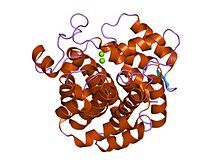
ADP-ribosylhydrolase
| ADP-ribosylhydrolase | |||||||||
|---|---|---|---|---|---|---|---|---|---|
 crystal structure of ribosylglycohydrolase mj1187 from methanococcus jannaschii | |||||||||
| Identifiers | |||||||||
| Symbol | ARH | ||||||||
| Pfam | PF03747 | ||||||||
| InterPro | IPR005502 | ||||||||
| SCOP2 | 1t5j / SCOPe / SUPFAM | ||||||||
| |||||||||
In molecular biology, the (ADP-ribosyl)hydrolase (ARH) family contains enzymes which catalyses the hydrolysis of ADP-ribosyl modifications from proteins, nucleic acids and small molecules.[1]
Types
[edit]This family has three members in humans (ARH1-3): ARH1, also termed [Protein ADP-ribosylarginine] hydrolase, cleaves ADP-ribose-L-arginine,[2] ARH2, which is predicted to be enzymatically inactive,[3] and ARH3, which cleaves primarily ADP-ribose-L-serine, but was shown to also hydrolyse poly(ADP-ribose), 1''-O-acetyl-ADP-ribose and alpha-nicotinamide adenine dinucleotide.[4][5][6][7] The family also includes ADP-ribosyl-(dinitrogen reductase) hydrolase (DraG) known to regulate dinitrogenase reductase, a key enzyme of the nitrogen fixating pathway in bacteria,[8][1] and most surprisingly jellyfish crystallins,[8][9] although the latter proteins appear to have lost the presumed active site residues.
| Class | Species | Intracellular location |
Activity | Function | ||
|---|---|---|---|---|---|---|
| Bacteria | Human | Others | ||||
| I | ARH1 | endoplasmic reticulum, cytoplasm | ADP-ribosylarginine hydrolase | inflammation, genomic stability | ||
| II | ARH2 | cytoplasm, cardiac sarcomeres | inactive | heart chamber outgrowth | ||
| III | ARH3 | Nucleus, cytoplasm | ADP-ribosylserine hydrolase | DNA repair | ||
| IV | Crystallin J1[9] and SelJ[10] | inactive | Crystallin | |||
| V | DraG | ADP-ribosylarginine hydrolase | Regulation of nitrogen fixation | |||
See also
[edit]References
[edit]- ^ a b Rack JG, Palazzo L, Ahel I (March 2020). "(ADP-ribosyl)hydrolases: structure, function, and biology". Genes & Development. 34 (5–6): 263–284. doi:10.1101/gad.334631.119. PMC 7050489. PMID 32029451.
- ^ Takada T, Iida K, Moss J (August 1993). "Cloning and site-directed mutagenesis of human ADP-ribosylarginine hydrolase". The Journal of Biological Chemistry. 268 (24): 17837–43. doi:10.1016/S0021-9258(17)46780-9. PMID 8349667.
- ^ Smith SJ, Towers N, Saldanha JW, Shang CA, Mahmood SR, Taylor WR, Mohun TJ (August 2016). "The cardiac-restricted protein ADP-ribosylhydrolase-like 1 is essential for heart chamber outgrowth and acts on muscle actin filament assembly". Developmental Biology. 416 (2): 373–88. doi:10.1016/j.ydbio.2016.05.006. PMC 4990356. PMID 27217161.
- ^ Fontana P, Bonfiglio JJ, Palazzo L, Bartlett E, Matic I, Ahel I (June 2017). "Serine ADP-ribosylation reversal by the hydrolase ARH3". eLife. 6: e28533. doi:10.7554/eLife.28533. PMC 5552275. PMID 28650317.
- ^ Stevens LA, Kato J, Kasamatsu A, Oda H, Lee DY, Moss J (December 2019). "The ARH and Macrodomain Families of α-ADP-ribose-acceptor Hydrolases Catalyze α-NAD + Hydrolysis". ACS Chemical Biology. 14 (12): 2576–2584. doi:10.1021/acschembio.9b00429. PMC 8388552. PMID 31599159.
- ^ Ono T, Kasamatsu A, Oka S, Moss J (November 2006). "The 39-kDa poly(ADP-ribose) glycohydrolase ARH3 hydrolyzes O-acetyl-ADP-ribose, a product of the Sir2 family of acetyl-histone deacetylases". Proceedings of the National Academy of Sciences of the United States of America. 103 (45): 16687–91. Bibcode:2006PNAS..10316687O. doi:10.1073/pnas.0607911103. PMC 1636516. PMID 17075046.
- ^ Oka S, Kato J, Moss J (January 2006). "Identification and characterization of a mammalian 39-kDa poly(ADP-ribose) glycohydrolase". The Journal of Biological Chemistry. 281 (2): 705–13. doi:10.1074/jbc.M510290200. PMID 16278211. S2CID 19256217.
- ^ a b Fitzmaurice WP, Saari LL, Lowery RG, Ludden PW, Roberts GP (August 1989). "Genes coding for the reversible ADP-ribosylation system of dinitrogenase reductase from Rhodospirillum rubrum". Molecular & General Genetics. 218 (2): 340–7. doi:10.1007/BF00331287. PMID 2506427. S2CID 35664554.
- ^ a b Piatigorsky J, Horwitz J, Norman BL (June 1993). "J1-crystallins of the cubomedusan jellyfish lens constitute a novel family encoded in at least three intronless genes". The Journal of Biological Chemistry. 268 (16): 11894–901. doi:10.1016/S0021-9258(19)50284-8. PMID 8505315.
- ^ Castellano S, Lobanov AV, Chapple C, Novoselov SV, Albrecht M, Hua D, et al. (November 2005). "Diversity and functional plasticity of eukaryotic selenoproteins: identification and characterization of the SelJ family". Proceedings of the National Academy of Sciences of the United States of America. 102 (45): 16188–93. doi:10.1073/pnas.0505146102. PMC 1283428. PMID 16260744.
