File:ZhaopingZhe2015Fig1 pcbi.1004375.g001.jpg
Page contents not supported in other languages.
Tools
Actions
General
In other projects
Appearance
ZhaopingZhe2015Fig1_pcbi.1004375.g001.jpg (700 × 396 pixels, file size: 69 KB, MIME type: image/jpeg)
| This is a file from the Wikimedia Commons. Information from its description page there is shown below. Commons is a freely licensed media file repository. You can help. |
Summary
| DescriptionZhaopingZhe2015Fig1 pcbi.1004375.g001.jpg |
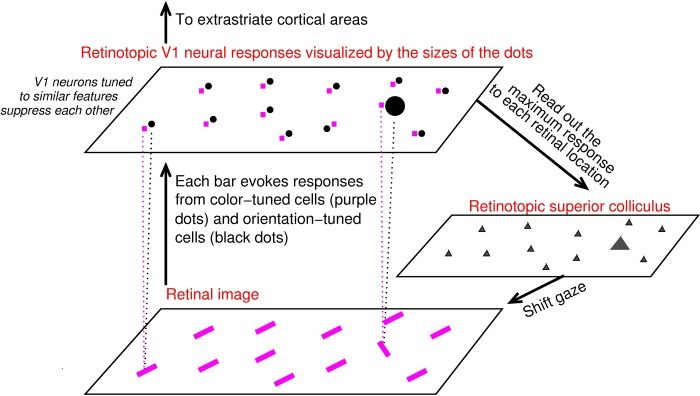
English: V1 saliency hypothesis states that the bottom-up saliency of a location is represented by the maximum V1 response to this location.In this schematic, V1 is simplified to contain only two kinds of neurons, one tuned to color (their responses are visualized by the purple dots) and the other tuned to orientation (black dots). Each input bar evokes responses in a cell tuned to its color and another cell tuned to its orientation (indicated for two input bars by linking each bar to its two evoked responses by dotted lines), and the receptive fields of these two cells cover the same bar location even though (for better visualization) the dots representing these cells are not overlapping in the cortical map. Iso-feature suppression makes nearby V1 neurons tuned to similar features (e.g., similar color or similar orientation) suppress each other. The orientation singleton in this image evokes the highest V1 response to this image because the orientation-tuned neuron responding to it escapes iso-orientation suppression. The color tuned neuron tuned and responding to the singleton’s color is under iso-color suppression. The saliency map is likely read out by the superior colliculus to execute gaze shifts to salient locations (see Zhaoping L. Understanding Vision: theory, models, and data. Oxford University Press; 2014.) |
| Date | The paper containing this figure was published online 2015 Oct 6 |
| Source | Own work |
| Author | Zhaopingli |
Licensing
I, the copyright holder of this work, hereby publish it under the following license:
This file is licensed under the Creative Commons Attribution-Share Alike 4.0 International license.
- You are free:
- to share – to copy, distribute and transmit the work
- to remix – to adapt the work
- Under the following conditions:
- attribution – You must give appropriate credit, provide a link to the license, and indicate if changes were made. You may do so in any reasonable manner, but not in any way that suggests the licensor endorses you or your use.
- share alike – If you remix, transform, or build upon the material, you must distribute your contributions under the same or compatible license as the original.
Captions
This explains a proposed function of the primary visual cortex, from Fig. 1 of a publication https://www.ncbi.nlm.nih.gov/pmc/articles/PMC4595278/
Items portrayed in this file
depicts
image/jpeg
70,190 byte
396 pixel
700 pixel
fa68037b846a35b0f13dc630d4063b733b2968d3
File history
Click on a date/time to view the file as it appeared at that time.
| Date/Time | Thumbnail | Dimensions | User | Comment | |
|---|---|---|---|---|---|
| current | 07:36, 9 May 2020 |  | 700 × 396 (69 KB) | Zhaopingli | Uploaded own work with UploadWizard |
File usage
The following 2 pages use this file:
Metadata
This file contains additional information, probably added from the digital camera or scanner used to create or digitize it.
If the file has been modified from its original state, some details may not fully reflect the modified file.
| Image title |
|
|---|---|
| Publisher | Public Library of Science |
| Short title |
|
| Copyright holder |
|
| Source | info:doi/10.1371/journal.pcbi.1004375 |
| Date(s) | 6 October 2015 |
| Identifier | info:doi/info:doi/10.1371/journal.pcbi.1004375.g001 |

