Jump to content

File:Tensor tympani-muscle.jpg

Page contents not supported in other languages.
This is a file from the Wikimedia Commons
From Wikipedia, the free encyclopedia

Tensor_tympani-muscle.jpg (258 × 303 pixels, file size: 11 KB, MIME type: image/jpeg)

Summary

Description
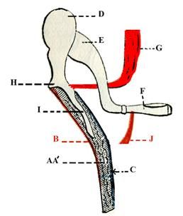
English: this figure allows to see the correct insertion of the tensor tempani muscle. AA’ ( two fibrous collagenic layers); B épidermis; C mucous membrane; D head of malleus; E incus; F stapes; G tensor tympani; H lateral process of malleus; I Manubrium of malleus; J stapes muscle.
Date
Source Tillaux Paul Jules, Traité d’Anatomie topographique avec applications à la chirurgie, Paris Asselin et Houzeau publishers (4°ed. 1884, p.125 ).
Author Tillaux Paul Jules (http://en.wikipedia.org/wiki/Paul_Jules_Tillaux => 1834-1904)

Licensing

Public domain

This work is in the public domain in its country of origin and other countries and areas where the copyright term is the author's life plus 100 years or fewer.


You must also include a United States public domain tag to indicate why this work is in the public domain in the United States.
This file has been identified as being free of known restrictions under copyright law, including all related and neighboring rights.

Captions

Add a one-line explanation of what this file represents

Items portrayed in this file

depicts

12 August 2011

File history

Click on a date/time to view the file as it appeared at that time.

Date/TimeThumbnailDimensionsUserComment
current19:47, 12 August 2011Thumbnail for version as of 19:47, 12 August 2011258 × 303 (11 KB)Auriol

The following page uses this file:

Global file usage

The following other wikis use this file:

Metadata