Jump to content

File:RLS 12blauLeg.png

Page contents not supported in other languages.
This is a file from the Wikimedia Commons
From Wikipedia, the free encyclopedia

Original file (883 × 1,332 pixels, file size: 1.56 MB, MIME type: image/png)

Summary

Description
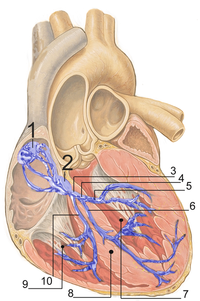
English: Electrical conduction system of the heart:
  1. Sinoatrial node
  2. Atrioventricular node
  3. Bundle of His
  4. Left bundle branch
  5. Left posterior fascicle
  6. Left-anterior fascicle
  7. Left ventricle
  8. Ventricular septum
  9. Right ventricle
  10. Right bundle branch
Français : Répartition du tissus nodal dans le coeur:
  1. nœud sinusal
  2. nœud atrio-ventriculaire
  3. faisceau de His
  4. branche gauche
  5. Faisceau postérieur gauche
  6. Faisceau antérieur gauche
  7. ventricule gauche
  8. Septum interventriculaire
  9. ventricule droit
  10. branche droite
Русский: Проводящая система сердца:
  1. Синоатриальный узел
  2. Атриовентрикулярный узел
  3. Пучок Гиса
  4. Левая ножка пучка Гиса
  5. Левая передняя ветвь
  6. Левая задняя ветвь
  7. Левый желудочек
  8. Межжелудочковая перегородка
  9. Правый желудочек
  10. Правая ножка пучка Гиса
Español: Sistema de conducción del corazón:
  1. Nódulo sinoauricular
  2. Nódulo auriculoventricular
  3. Haz de His
  4. Rama izquierda
  5. Fascículo posterior izquierdo
  6. Fascículo anterior izquierdo
  7. Ventrículo izquierdo
  8. Tabique interventricular
  9. Ventrículo derecho
  10. Rama derecha
Deutsch: Erregungsleitungssystem:
  1. Sinusknoten
  2. AV-Knoten
  3. His-Bündel
  4. Linker Tawara-Schenkel
  5. Linksposteriorer Faszikel
  6. Linksanteriorer Faszikel
  7. Linker Ventrikel
  8. Ventrikelseptum
  9. Rechter Ventrikel
  10. Rechter Tawara-Schenkel
Polski: Układ bodźcotwórczo-przewodzący:
  1. węzeł zatokowo-przedsionkowy
  2. węzeł przedsionkowo-komorowy
  3. pęczek Hisa
  4. lewa odnoga pęczka Hisa
  5. wiązka tylna lewej odnogi
  6. wiązka przednia lewej odnogi
  7. lewa komora
  8. przegroda międzykomorowa
  9. prawa komora
  10. prawa odnoga pęczka Hisa
Date
Source self made, based upon Image:Heart anterior view coronal section.jpg by Patrick J. Lynch (Patrick J. Lynch; illustrator; C. Carl Jaffe; MD; cardiologist Yale University Center for Advanced Instructional Media )
Author J. Heuser
Permission
(Reusing this file)
Creative Commons Attribution 2.5 License 2007

preserve our creative credits: Patrick J. Lynch, medical illustrator; C. Carl Jaffe, MD, cardiologist. https://creativecommons.org/licenses/by/2.5/

Licensing

w:en:Creative Commons
attribution
This file is licensed under the Creative Commons Attribution 2.5 Generic license.
You are free:
  • to share – to copy, distribute and transmit the work
  • to remix – to adapt the work
Under the following conditions:
  • attribution – You must give appropriate credit, provide a link to the license, and indicate if changes were made. You may do so in any reasonable manner, but not in any way that suggests the licensor endorses you or your use.

Captions

Add a one-line explanation of what this file represents

Items portrayed in this file

depicts

2 March 2007

File history

Click on a date/time to view the file as it appeared at that time.

Date/TimeThumbnailDimensionsUserComment
current17:51, 2 March 2007Thumbnail for version as of 17:51, 2 March 2007883 × 1,332 (1.56 MB)JHeuser{{Information |Description = Heart; conduction system |Source = self made, based upon Image:Heart anterior view coronal section.jpg by Patrick J. Lynch (Patrick J. Lynch; illustrator; C. Carl Jaffe; MD; cardiologist Yale University Center for Advanc

Global file usage

The following other wikis use this file: