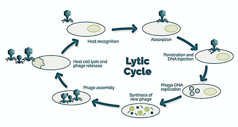
English: Lytic cycle
The process of the lytic cycle is divided into five steps.
First of all, the virus has to find and attach to the surface of the target bacteria. This attachment depends on the presence of specific receptors on the cell surface and determines the specificity and host range of a given bacteriophage. Different bacteriophages have different strategies or target molecules.
Most bacteriophages use cell wall molecules as receptors. These are known as somatic bacteriophages. Myoviridae, Siphoviridae, Podoviridae and Microviridae belong to this group. Another strategy is to use cell appendixes like F pili or flagella. These are called F-specific bacteriophages, which use the F or sexual pili as receptors and as a channel for "injecting" their nucleic acid. The lnoviridae and Leviviridae belong to this group.
There is no active positive attraction (positive taxis) between phages and host bacteria and consequently meeting of bacteria and phages depends on random encounters. Whether these encounters occur or not depends to a great extent on the concentrations of bacteria and bacteriophages. Some studies performed with somatic coliphages indicate that the sum of log10 concentrations of phages and host bacteria per ml that guarantee replication is around 6.5 (Jofre, 2009).
The second step is penetration, which consists of the injection of the viral genome and seldom molecules contained inside the capsid into the bacterial cytoplasm. This can happen through the cell wall (somatic bacteriophages) or through an appendage (flagella and the sexual pili).
Then the viral genome, using the metabolic tools of the host cell, starts replicating and the production of viral proteins takes place. This step requires the host to be metabolically active
When feces exit the gut, the change of environment causes a stress on the great majority of fecal bacteria, including coliform bacteria, that leads first to metabolic inactivity and then to death. This, together with the difficulties for viruses to come across a ceLL in a liquid matrix with a relatively poor concentration of phages and bacteria, makes viral reproduction outside the gut barely possible (Jofre, 2009).
The fourth step is the assembly of the different proteins and the genome, forming the capsomers that bind with each other and with the genetic material to build the capsid, and hence producing newborn virions which become visible (by electron microscopy) inside the bacterial cytoplasm.
Finally, the bacterial cell breaks, in a process known as Lysis, due to enzymatic action of some proteins coded in the viral genome, and new bacteriophages are released to the media. Some bacteriophages, such as for example M13, do not Lyse the cell, but this seems to be exceptional.
All this process can happen in Less than 30 minutes, and at the end between 100 and 200 (depending on the bacteriophage) new infectious phage particles are released.
- Jofre, J. (2009). "Is the replication of somatic coLiphages in water environments significant?" J. AppL. Microbiol, 106: 1059–1069.


