Jump to content

File:Figure1-micropublish4.jpg

Page contents not supported in other languages.
This is a file from the Wikimedia Commons
From Wikipedia, the free encyclopedia

Original file (5,500 × 3,332 pixels, file size: 2.97 MB, MIME type: image/jpeg)

Summary

Description
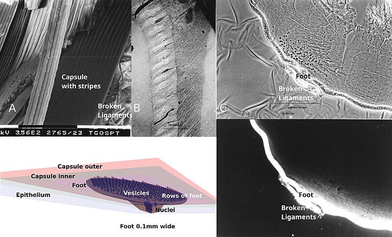
English: (A) Scanning electron micrograph of a micro-dissected sheep lens from a lamb less than a year old. Cells pull apart cleanly with membranes predominantly left intact. The capsule is very thin here, probably less than 2µm, as shown by the sheared edge being rounded but still appearing thin. The striped pattern near where the ligaments attach to the lens were not seen in large areas of well preserved thicker capsule elsewhere in the lens. Magnification bar 100µm (B) Transmission electron micrograph of lens capsule removed from a mouse, fixed and stained with heavy metals prior to ultra-thin sectioning. Top right is a copper supporting grid bar with adjacent darkly stained elongated cell nuclei. Circular white vesicles surround the nuclei and extend to the left and down into a band of the grayer cytoplasm. Further to the left is a nominally 10µm thick more lightly stained basement membrane with cytoplasmic feet and fibrils extending into and through it. (C) Interference contrast microscopy above and FITC coupled WGA fluorescence microscopy below can show structural features that parallel structures seen with electron microscopy. (D) A 3D highly stylized foot-feet model of basement membrane growth in a nominally 0.8mm diameter spherical 8 week old mouse lens based on Fig. 1C. The cells may form the foot from either the epithelium or the cells migrating along the ligaments towards the lens surface where they move to areas where they are needed. Blue spheres represent nuclei inside a cell. Smaller white spheres represent vesicles. The rows of numerous feet are highlighted by making them darker than the surrounding gray foot.
Date
Source Own work
Author Tgru001

Licensing

I, the copyright holder of this work, hereby publish it under the following license:
w:en:Creative Commons
attribution
This file is licensed under the Creative Commons Attribution 4.0 International license.
You are free:
  • to share – to copy, distribute and transmit the work
  • to remix – to adapt the work
Under the following conditions:
  • attribution – You must give appropriate credit, provide a link to the license, and indicate if changes were made. You may do so in any reasonable manner, but not in any way that suggests the licensor endorses you or your use.

Captions

Micrographs and 3D drawing of a "foot" structures on eye lens capsules

Items portrayed in this file

depicts

4 April 2023

image/jpeg

File history

Click on a date/time to view the file as it appeared at that time.

Date/TimeThumbnailDimensionsUserComment
current03:31, 2 August 2024Thumbnail for version as of 03:31, 2 August 20245,500 × 3,332 (2.97 MB)Tgru001Updated diagram to better reflect micrographs in the same image.
04:52, 24 April 2024Thumbnail for version as of 04:52, 24 April 20245,500 × 3,332 (8.48 MB)Tgru001Uploaded own work with UploadWizard

The following page uses this file:

Metadata