Jump to content

File:Endo Fracture.jpg

Page contents not supported in other languages.
This is a file from the Wikimedia Commons
From Wikipedia, the free encyclopedia

Original file (1,061 × 901 pixels, file size: 303 KB, MIME type: image/jpeg)

Summary

Description
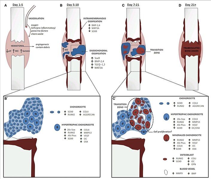
English: Endochondral Fracture Schematic: A fracture that is not rigidly stabilized will heal through a combination of intramembranous and endochondral ossification. Fracture healing progression: bone is depicted in red and cartilage in blue, time course corresponds to healing in a mouse tibia. (A) The first stage of fracture healing is formation of a hematoma to initiate an inflammatory cascade characterized in part by an abundance of pro-inflammatory M1 macrophages, low pH, and elevated lactate level. (B) Next, local osteochondral progenitors differentiate into bone and cartilage to initiate healing. Intramembranous ossification occurs primarily at the periosteum and endosteum of the fracture callus, while the soft cartilage callus forms in the central portion of the fracture where there is maximal mobility. For healing to progress normally, inflammation must be down-regulated and there is a shift in the macrophage population from a pro-inflammatory M1 state to an anti-inflammatory M2 state. (B’) Within the cartilage callus chondrocytes differentiate and mature in a parallel fashion to the growth plate. (C) Conversion of cartilage to bone during endochondral ossification occurs concomitantly with the invasion of blood vessels. (C’) Blood vessels lead to mineralization of the cartilage matrix and new data suggest that these cells transform directly into osteoblasts. Chondrocytes do not undergo significant apoptosis and may re-enter the cell cycle. (D) The cartilage callus becomes fully converted to a trabeculated bone that will bridge the full fracture defect. This trabeculated structure will be remodeled into a cortical bone that is almost indistinguishable in form and function from the native bone.
Date
Source https://www.ncbi.nlm.nih.gov/pmc/articles/PMC4318416/
Author Chelsea S. Bahney, Diane P. Hu, Theodore Miclau, III, and Ralph S. Marcucio

Licensing

w:en:Creative Commons
attribution
This file is licensed under the Creative Commons Attribution 4.0 International license.
You are free:
  • to share – to copy, distribute and transmit the work
  • to remix – to adapt the work
Under the following conditions:
  • attribution – You must give appropriate credit, provide a link to the license, and indicate if changes were made. You may do so in any reasonable manner, but not in any way that suggests the licensor endorses you or your use.

Captions

Endochondral Fracture Schematic

Items portrayed in this file

depicts

2 February 2024

File history

Click on a date/time to view the file as it appeared at that time.

Date/TimeThumbnailDimensionsUserComment
current01:52, 2 February 2024Thumbnail for version as of 01:52, 2 February 20241,061 × 901 (303 KB)PecMoUploaded a work by Chelsea S. Bahney, Diane P. Hu, Theodore Miclau, III, and Ralph S. Marcucio from https://www.ncbi.nlm.nih.gov/pmc/articles/PMC4318416/ with UploadWizard

The following page uses this file:

Global file usage

The following other wikis use this file:

Metadata