Jump to content

File:Dilophosaurus pathologies.PNG

Page contents not supported in other languages.
This is a file from the Wikimedia Commons
From Wikipedia, the free encyclopedia

Original file (4,066 × 5,220 pixels, file size: 11.94 MB, MIME type: image/png)

Description

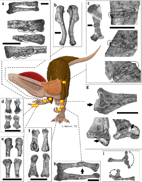
Pathological features in the forelimbs and left scapula of UCMP 37302 (Dilophosaurus wetherilli).

(a) Right radius and ulna (above) and enlargements of distal end of radius (below) in (from top to bottom) lateral, abductor, and medial views; broken outline indicates three bony tumors. (b) Left and right humerus (left humerus on left, right humerus on right) in lateral view, each photographed with lateral epicondyle directly facing the viewer, with heavy broken line indicating the midline of the posterior (retractor) surface of each to show the abnormal degree of torsion in the right humerus. (c) Medial surface of left scapula, with broken outline indicating fracture. (d) Left (on left) and right (on right) manual phalanx III-1 in dorsal (top) and palmar (bottom) views, with broken lines indicating plane of articulation with adjacent bones, to show the alteration of this plane in the right-hand phalanx. (e) Distal ends of left (on left) and right (on right) metacarpal III in lateral/abductor view (top) and palmar view (bottom), with broken outline indicating edge of articular surface, to show abnormal truncation of articular surface in right metacarpal III. (f) Left manual phalanx I-1 (on left), with its right-hand counterpart for comparison (on right), in palmar (top) and lateral/abductor (bottom) views, with broken outlines indicating healed fibriscesses. (g) Medial surface of left ulna, with broken outline indicating healed fibriscess and arrow indicating abnormal bony growth. (h) Left radius and ulna in medial view, with arrow indicating healed fracture. (i) Left (top) and right (bottom) metacarpal III and phalanx III-1, with phalanx III-1 in full extension and full flexion, to show the reduced range of motion of this digit in the right hand. Scale bars = 50 mm.
Date
Source http://journals.plos.org/plosone/article?id=10.1371/journal.pone.0149140
Author Leandra Walters, Phil Senter, Sara L. Juengst
w:en:Creative Commons
attribution
This file is licensed under the Creative Commons Attribution 2.5 Generic license.
You are free:
  • to share – to copy, distribute and transmit the work
  • to remix – to adapt the work
Under the following conditions:
  • attribution – You must give appropriate credit, provide a link to the license, and indicate if changes were made. You may do so in any reasonable manner, but not in any way that suggests the licensor endorses you or your use.
PLOS
PLOS
This file was published in a Public Library of Science journal. Their website states that the content of all PLOS journals is published under the Creative Commons Attribution 4.0 license (or its previous version depending on the publication date), unless indicated otherwise.

Captions

Add a one-line explanation of what this file represents

Items portrayed in this file

depicts

12,520,597 byte

5,220 pixel

4,066 pixel

image/png

993aee8fc6e0ae50e0152e3dd1ce9cab3a8c9748

File history

Click on a date/time to view the file as it appeared at that time.

Date/TimeThumbnailDimensionsUserComment
current17:33, 25 February 2016Thumbnail for version as of 17:33, 25 February 20164,066 × 5,220 (11.94 MB)FunkMonk{{Information |Description=Pathological features in the forelimbs and left scapula of UCMP 37302 (Dilophosaurus wetherilli). (a) Right radius and ulna (above) and enlargements of distal end of radius (below) in (from top to bottom) lateral, abductor,...

Global file usage

The following other wikis use this file:

Metadata