
Ehlers–Danlos syndrome: Difference between revisions
m Corrected information that was summarized inaccurately |
Ira Leviton (talk | contribs) m Fixed a reference. Please see Category:CS1 errors: dates. |
||
| (46 intermediate revisions by 37 users not shown) | |||
| Line 1: | Line 1: | ||
{{short description|Group of genetic connective tissues disorders}} |
{{short description|Group of genetic connective tissues disorders}} |
||
{{cs1 config|name-list-style=vanc}} |
{{cs1 config|name-list-style=vanc}} |
||
{{Infobox medical condition |
{{Infobox medical condition |
||
| name = Ehlers–Danlos syndrome |
| name = Ehlers–Danlos syndrome |
||
| image = PMC3504533 1471-2415-12-47-2 (cropped).png |
| image = PMC3504533 1471-2415-12-47-2 (cropped).png |
||
| alt = |
| alt = |
||
| caption = Individual with EDS displaying skin hyperelasticity |
| caption = Individual with classical EDS displaying skin hyperelasticity |
||
| field = [[Medical genetics]] |
| field = [[Medical genetics]] |
||
| pronounce = {{IPAc-en|ˈ|eɪ|l|ər|z|_|ˈ|d|æ|n|l|ɒ|s}} |
| pronounce = {{IPAc-en|ˈ|eɪ|l|ər|z|_|ˈ|d|æ|n|l|ɒ|s}} |
||
| Line 25: | Line 25: | ||
}} |
}} |
||
<!-- Definition and symptoms --> |
<!-- Definition and symptoms --> |
||
'''Ehlers–Danlos syndromes''' ('''EDS''') are a group of 13 [[genetic disorders|genetic]] [[connective tissue disease|connective-tissue disorders]].<ref>{{cite journal | vauthors = Dattagupta A, Williamson S, El Nihum LI, Petak S | title = A Case of Spondylodysplastic Ehlers–Danlos Syndrome With Comorbid Hypophosphatasia | journal = AACE Clinical Case Reports | volume = 8 | issue = 6 | pages = 255–258 | date = 2022-11-01 | pmid = 36447830 | pmc = 9701907 | doi = 10.1016/j.aace.2022.08.005}}</ref> Symptoms often include loose joints, joint pain, stretchy velvety skin, and abnormal scar formation.<ref name="NIHGHR2016" /> These may be noticed at birth or in early childhood.<ref name="Net2012">{{cite book| vauthors = Anderson BE |title=The Netter Collection of Medical Illustrations – Integumentary System |via=E-Book|date=2012|publisher=Elsevier Health Sciences|isbn=978-1455726646 |page=235 |edition=2nd |url=https://books.google.com/books?id=LOBYSIiRL8oC&pg=PA235|url-status=live |archive-url=https://web.archive.org/web/20171105195522/https://books.google.com/books?id=LOBYSIiRL8oC&pg=PA235|archive-date=2017-11-05}}</ref> Complications may include [[aortic dissection]], [[joint dislocations]], [[scoliosis]], [[chronic pain]], or early [[osteoarthritis]].<ref name=NIHGHR2016/><ref name=Law2005/> The current classification was last updated in 2017, when a number of rarer forms of EDS were added.<ref name="NIHGHR2016" /> |
'''Ehlers–Danlos syndromes''' ('''EDS''') are a group of 13 [[genetic disorders|genetic]] [[connective tissue disease|connective-tissue disorders]].<ref>{{cite journal | vauthors = Dattagupta A, Williamson S, El Nihum LI, Petak S | title = A Case of Spondylodysplastic Ehlers–Danlos Syndrome With Comorbid Hypophosphatasia | journal = AACE Clinical Case Reports | volume = 8 | issue = 6 | pages = 255–258 | date = 2022-11-01 | pmid = 36447830 | pmc = 9701907 | doi = 10.1016/j.aace.2022.08.005}}</ref> Symptoms often include loose joints, joint pain, stretchy velvety skin, and abnormal scar formation.<ref name="NIHGHR2016" /> These may be noticed at birth or in early childhood.<ref name="Net2012">{{cite book| vauthors = Anderson BE |title=The Netter Collection of Medical Illustrations – Integumentary System |via=E-Book|date=2012|publisher=Elsevier Health Sciences|isbn=978-1455726646 |page=235 |edition=2nd |url=https://books.google.com/books?id=LOBYSIiRL8oC&pg=PA235|url-status=live |archive-url=https://web.archive.org/web/20171105195522/https://books.google.com/books?id=LOBYSIiRL8oC&pg=PA235|archive-date=2017-11-05}}</ref> Complications may include [[aortic dissection]], [[joint dislocations]], [[scoliosis]], [[chronic pain]], or early [[osteoarthritis]].<ref name=NIHGHR2016/><ref name=Law2005/> The current classification was last updated in 2017, when a number of rarer forms of EDS were added.<ref name="NIHGHR2016" /><ref name=":1">{{cite journal | vauthors = Malfait F, Francomano C, Byers P, Belmont J, Berglund B, Black J, Bloom L, Bowen JM, Brady AF, Burrows NP, Castori M, Cohen H, Colombi M, Demirdas S, De Backer J, De Paepe A, Fournel-Gigleux S, Frank M, Ghali N, Giunta C, Grahame R, Hakim A, Jeunemaitre X, Johnson D, Juul-Kristensen B, Kapferer-Seebacher I, Kazkaz H, Kosho T, Lavallee ME, Levy H, Mendoza-Londono R, Pepin M, Pope FM, Reinstein E, Robert L, Rohrbach M, Sanders L, Sobey GJ, Van Damme T, Vandersteen A, van Mourik C, Voermans N, Wheeldon N, Zschocke J, Tinkle B | display-authors = 6 | title = The 2017 international classification of the Ehlers–Danlos syndromes | journal = American Journal of Medical Genetics. Part C, Seminars in Medical Genetics | volume = 175 | issue = 1 | pages = 8–26 | date = March 2017 | pmid = 28306229 | doi = 10.1002/ajmg.c.31552 | s2cid = 4440499 | doi-access = free}} {{free access}}</ref> |
||
<!-- Etiopathogenesis --> |
<!-- Etiopathogenesis --> |
||
| Line 34: | Line 34: | ||
<!-- Treatment and prognosis --> |
<!-- Treatment and prognosis --> |
||
A cure is not yet known<ref name=Fer2016>{{cite book| vauthors = Ferri FF |title=Ferri's Netter Patient Advisor |date=2016 |publisher=Elsevier Health Sciences |isbn=9780323393249 |page=939|url=https://books.google.com/books?id=Dz_dCwAAQBAJ&pg=PA939|url-status=live |archive-url= https://web.archive.org/web/20171105195522/https://books.google.com/books?id=Dz_dCwAAQBAJ&pg=PA939 |archive-date=2017-11-05}}</ref> and treatment is [[supportive treatment|supportive]] in nature.<ref name=Law2005/> [[Physical therapy]] and bracing may help strengthen muscles and support joints.<ref name=Law2005>{{cite journal | vauthors = Lawrence EJ | title = The clinical presentation of Ehlers–Danlos syndrome | journal = Advances in Neonatal Care | volume = 5 | issue = 6 | pages = 301–314 | date = December 2005 | pmid = 16338669 | doi = 10.1016/j.adnc.2005.09.006 | s2cid = 7717730}}</ref> Some forms of EDS result in a normal [[life expectancy]], but those that affect [[blood vessel]]s generally decrease it.<ref name=Fer2016/> All forms of EDS can result in fatal outcomes for some patients.<ref name="auto">{{cite journal | vauthors = Brady AF, Demirdas S, Fournel-Gigleux S, Ghali N, Giunta C, Kapferer-Seebacher I, Kosho T, Mendoza-Londono R, Pope MF, Rohrbach M, Van Damme T, Vandersteen A, van Mourik C, Voermans N, Zschocke J, Malfait F | display-authors = 6 | title = The Ehlers–Danlos syndromes, rare types | journal = American Journal of Medical Genetics. Part C, Seminars in Medical Genetics | volume = 175 | issue = 1 | pages = 70–115 | date = March 2017 | pmid = 28306225 | doi = 10.1002/ajmg.c.31550 | s2cid = 4439633 | doi-access = free}}</ref><ref name="auto2">{{cite journal | vauthors = Doolan BJ, Lavallee ME, Hausser I, Schubart JR, Michael Pope F, Seneviratne SL, Winship IM, Burrows NP | display-authors = 6 | title = Extracutaneous features and complications of the Ehlers–Danlos syndromes: A systematic review | journal = Frontiers in Medicine | volume = 10 | pages = 1053466 | date = 23 Jan 2023 | pmid = 36756177 | pmc = 9899794 | doi = 10.3389/fmed.2023.1053466 | doi-access = free}}</ref><ref name="pmid35986728">{{cite journal | vauthors = Marathe N, Lohkamp LN, Fehlings MG | title = Spinal manifestations of Ehlers–Danlos syndrome: a scoping review | journal = Journal of Neurosurgery. Spine | volume = 37 | issue = 6 | pages = 783–793 | date = December 2022 | pmid = 35986728 | pmc = | doi = 10.3171/2022.6.SPINE211011 | s2cid = 251694109}}</ref> |
A cure is not yet known,<ref name=Fer2016>{{cite book| vauthors = Ferri FF |title=Ferri's Netter Patient Advisor |date=2016 |publisher=Elsevier Health Sciences |isbn=9780323393249 |page=939|url=https://books.google.com/books?id=Dz_dCwAAQBAJ&pg=PA939|url-status=live |archive-url= https://web.archive.org/web/20171105195522/https://books.google.com/books?id=Dz_dCwAAQBAJ&pg=PA939 |archive-date=2017-11-05}}</ref> and treatment is [[supportive treatment|supportive]] in nature.<ref name=Law2005/> [[Physical therapy]] and bracing may help strengthen muscles and support joints.<ref name=Law2005>{{cite journal | vauthors = Lawrence EJ | title = The clinical presentation of Ehlers–Danlos syndrome | journal = Advances in Neonatal Care | volume = 5 | issue = 6 | pages = 301–314 | date = December 2005 | pmid = 16338669 | doi = 10.1016/j.adnc.2005.09.006 | s2cid = 7717730}}</ref> Some forms of EDS result in a normal [[life expectancy]], but those that affect [[blood vessel]]s generally decrease it.<ref name=Fer2016/> All forms of EDS can result in fatal outcomes for some patients.<ref name="auto">{{cite journal | vauthors = Brady AF, Demirdas S, Fournel-Gigleux S, Ghali N, Giunta C, Kapferer-Seebacher I, Kosho T, Mendoza-Londono R, Pope MF, Rohrbach M, Van Damme T, Vandersteen A, van Mourik C, Voermans N, Zschocke J, Malfait F | display-authors = 6 | title = The Ehlers–Danlos syndromes, rare types | journal = American Journal of Medical Genetics. Part C, Seminars in Medical Genetics | volume = 175 | issue = 1 | pages = 70–115 | date = March 2017 | pmid = 28306225 | doi = 10.1002/ajmg.c.31550 | s2cid = 4439633 | doi-access = free}}</ref><ref name="auto2">{{cite journal | vauthors = Doolan BJ, Lavallee ME, Hausser I, Schubart JR, Michael Pope F, Seneviratne SL, Winship IM, Burrows NP | display-authors = 6 | title = Extracutaneous features and complications of the Ehlers–Danlos syndromes: A systematic review | journal = Frontiers in Medicine | volume = 10 | pages = 1053466 | date = 23 Jan 2023 | pmid = 36756177 | pmc = 9899794 | doi = 10.3389/fmed.2023.1053466 | doi-access = free}}</ref><ref name="pmid35986728">{{cite journal | vauthors = Marathe N, Lohkamp LN, Fehlings MG | title = Spinal manifestations of Ehlers–Danlos syndrome: a scoping review | journal = Journal of Neurosurgery. Spine | volume = 37 | issue = 6 | pages = 783–793 | date = December 2022 | pmid = 35986728 | pmc = | doi = 10.3171/2022.6.SPINE211011 | s2cid = 251694109}}</ref> |
||
<!-- Epidemiology and history --> |
<!-- Epidemiology and history --> |
||
| Line 41: | Line 41: | ||
== Types == |
== Types == |
||
In 2017, 13 subtypes of EDS were classified using specific diagnostic criteria.<ref name="GARD2017"/> According to the [[Ehlers–Danlos Society]], the syndromes can also be grouped by the symptoms determined by specific gene mutations. Group A disorders are those that affect primary collagen structure and processing. Group B disorders affect collagen folding and crosslinking. Group C are disorders of structure and function of myomatrix. Group D disorders are those that affect glycosaminoglycan biosynthesis. Group E disorders are characterized by defects in the complement pathway. Group F are disorders of intracellular processes, and Group G is considered to be unresolved forms of EDS.<ref name=":2">{{cite web|url=https://www.ehlers-danlos.com/eds-types/|title=The Types of EDS|website=The Ehlers Danlos Society|access-date=2019-11-06}}</ref> |
In 2017, 13 subtypes of EDS were classified using specific diagnostic criteria.<ref name="GARD2017"/> According to the [[Ehlers–Danlos Society]], the syndromes can also be grouped by the symptoms determined by specific gene mutations. Group A disorders are those that affect primary collagen structure and processing. Group B disorders affect collagen folding and crosslinking. Group C are disorders of structure and function of myomatrix. Group D disorders are those that affect [[glycosaminoglycan]] biosynthesis. Group E disorders are characterized by defects in the complement pathway. Group F are disorders of intracellular processes, and Group G is considered to be unresolved forms of EDS.<ref name=":2">{{cite web|url=https://www.ehlers-danlos.com/eds-types/|title=The Types of EDS|website=The Ehlers Danlos Society|access-date=2019-11-06}}</ref> |
||
===Hypermobile EDS (hEDS)=== |
===Hypermobile EDS (hEDS)=== |
||
Hypermobile EDS (hEDS, formerly categorized as type 3) is mainly characterized by hypermobility that affects both large and small joints. It may lead to frequent joint [[subluxations]] (partial dislocations) and dislocations. In general, people with this variant have skin that is soft, smooth, and velvety and bruises easily, and may have chronic muscle and/or bone pain.<ref name="GARD2017"/> It affects the skin less than other forms. It has no available genetic test.<ref name="Levy_2018"/> hEDS is the most common of the 19 types of connective tissue disorders. Since no genetic test exists, providers have to diagnose hEDS based on what they know about the condition and the patient's physical attributes. Other than the general signs, attributes can include faulty connective tissues throughout the body, musculoskeletal issues, and family history. Along with these general signs and side effects, patients can have trouble healing.<ref>{{cite web|url=https://www.ehlers-danlos.org/what-is-eds/information-on-eds/hypermobile-eds-and-hypermobility-spectrum-disorders/|title=Hypermobile EDS and Hypermobility Spectrum Disorders| vauthors = Carter K | work = Ehlers–Danlos Support UK}}</ref> |
Hypermobile EDS (hEDS, formerly categorized as type 3) is mainly characterized by hypermobility that affects both large and small joints. It may lead to frequent joint [[subluxations]] (partial dislocations) and dislocations. In general, people with this variant have skin that is soft, smooth, and velvety and bruises easily, and may have chronic muscle and/or bone pain.<ref name="GARD2017"/> It affects the skin less than other forms. It has no available genetic test.<ref name="Levy_2018"/> hEDS is the most common of the 19 types of connective tissue disorders. Since no genetic test exists, providers have to diagnose hEDS based on what they know about the condition and the patient's physical attributes. Other than the general signs, attributes can include faulty connective tissues throughout the body, musculoskeletal issues, and family history. Along with these general signs and side effects, patients can have trouble healing.<ref>{{cite web|url=https://www.ehlers-danlos.org/what-is-eds/information-on-eds/hypermobile-eds-and-hypermobility-spectrum-disorders/|title=Hypermobile EDS and Hypermobility Spectrum Disorders| vauthors = Carter K | work = Ehlers–Danlos Support UK}}</ref> |
||
Pregnant individuals who have hEDS are at an increased risk for complications. Some possible complications are pre-labor rupture of membranes, a drop in blood pressure with anesthesia, precipitate birth (very fast, active labor), malposition of the fetus, and increased bleeding. Individuals with hEDS may run the risk of falling, postpartum depression (more than the general population), and slow healing from the birthing process. |
Pregnant individuals who have hEDS are at an increased risk for complications. Some possible complications are pre-labor rupture of membranes, a drop in blood pressure with anesthesia, precipitate birth (very fast, active labor), malposition of the fetus, and increased bleeding. Individuals with hEDS may run the risk of falling, [[postpartum depression]] (more than the general population), and slow healing from the birthing process.<ref>{{cite web|url=https://www.ehlers-danlos.org/information/pregnancy-birth-feeding-and-hypermobile-ehlers-danlos-syndrome-hypermobility-spectrum-disorders/|title=Pregnancy, birth, feeding, and hypermobile Ehlers–Danlos syndrome/hypermobility spectrum disorders | work = The Ehlers–Danlos Support UK|access-date=2019-11-22}}</ref> |
||
The Medical University of South Carolina discovered a gene variant common with hEDS patients.<ref>{{Cite web | vauthors = Cantu L | date = 14 July 2021 |title=MUSC researchers announce gene mutation discovery associated with EDS |url=https://web.musc.edu/about/news-center/2021/07/14/musc-researchers-announce-gene-mutation-discovery-associated-with-eds-ehlers-danlos |access-date=2023-02-08 | work = Medical University of South Carolina}}</ref> |
The Medical University of South Carolina discovered a gene variant common with hEDS patients.<ref>{{Cite web | vauthors = Cantu L | date = 14 July 2021 |title=MUSC researchers announce gene mutation discovery associated with EDS |url=https://web.musc.edu/about/news-center/2021/07/14/musc-researchers-announce-gene-mutation-discovery-associated-with-eds-ehlers-danlos |access-date=2023-02-08 | work = Medical University of South Carolina}}</ref> |
||
| Line 54: | Line 54: | ||
While 12 of the 13 subtypes of EDS have genetic variations that can be tested for by [[genetic testing]], there is no known genetic cause of hEDS. Recently, several labs and research initiatives have been attempting to uncover a potential hEDS gene. In 2018, the [[Ehlers–Danlos Society]] began the Hypermobile Ehlers–Danlos Genetic Evaluation (HEDGE) study.<ref>{{cite web |url=https://www.ehlers-danlos.com/hedge/|title=HEDGE Study| website= ehlers-danlos.com| publisher= | date= | access-date=}}</ref> The ongoing study has screened over 1,000 people who have been diagnosed with hEDS by the 2017 criteria to evaluate their genome for a common mutation. To date, 200 people with hEDS have had [[whole genome sequencing]], and 500 have had whole [[Exome sequencing|exome]] sequencing; this study aims to increase those numbers significantly.{{citation needed|date=September 2022}} |
While 12 of the 13 subtypes of EDS have genetic variations that can be tested for by [[genetic testing]], there is no known genetic cause of hEDS. Recently, several labs and research initiatives have been attempting to uncover a potential hEDS gene. In 2018, the [[Ehlers–Danlos Society]] began the Hypermobile Ehlers–Danlos Genetic Evaluation (HEDGE) study.<ref>{{cite web |url=https://www.ehlers-danlos.com/hedge/|title=HEDGE Study| website= ehlers-danlos.com| publisher= | date= | access-date=}}</ref> The ongoing study has screened over 1,000 people who have been diagnosed with hEDS by the 2017 criteria to evaluate their genome for a common mutation. To date, 200 people with hEDS have had [[whole genome sequencing]], and 500 have had whole [[Exome sequencing|exome]] sequencing; this study aims to increase those numbers significantly.{{citation needed|date=September 2022}} |
||
Promising outcomes of this increased screening have been reported by the Norris Lab, led by Russell Norris, in the Department of Regenerative Medicine and Cell Biology at [[Medical University of South Carolina]].<ref>{{cite web |url=https://www.thenorrislab.com/home|title= About the Lab| publisher= Norris Lab, [[Medical University of South Carolina]]| date= | access-date=}}</ref> Using [[CRISPR]] Cas-9 mediated genome editing on mouse models of the disease, the lab has recently identified a "very strong candidate gene"<ref>{{cite journal | vauthors = Gensemer C, Burks R, Kautz S, Judge DP, Lavallee M, Norris RA | title = Hypermobile Ehlers–Danlos syndromes: Complex phenotypes, challenging diagnoses, and poorly understood causes | journal = Developmental Dynamics | volume = 250 | issue = 3 | pages = 318–344 | date = March 2021 | pmid = 32629534 | pmc = 7785693 | doi = 10.1002/dvdy.220}}</ref> for hEDS. This finding, and a greater understanding of cardiac complications associated with the majority of EDS subtypes, has led to the development of multiple druggable pathways involved in [[Aorta|aortic]] and [[mitral valve]] diseases. While this candidate gene has not been publicly identified, the Norris lab has conducted several studies involving small population genome sequencing and come up with a working list of possible hEDS genes. A mutation in [[Collagen, type III, alpha 1|''COL3A1'']]<ref>{{cite journal | vauthors = Narcisi P, Richards AJ, Ferguson SD, Pope FM | title = A family with Ehlers–Danlos syndrome type III/articular hypermobility syndrome has a glycine 637 to serine substitution in type III collagen | journal = Human Molecular Genetics | volume = 3 | issue = 9 | pages = 1617–1620 | date = September 1994 | pmid = 7833919 | doi = 10.1093/hmg/3.9.1617}}</ref> in a single family with autosomal dominant hEDS phenotype was found to cause reduced collagen secretion and an over-modification of collagen. In 35 families, copy number alterations in ''[[TPSAB1]]'',<ref>{{cite journal | vauthors = Lyons JJ, Yu X, Hughes JD, Le QT, Jamil A, Bai Y, Ho N, Zhao M, Liu Y, O'Connell MP, Trivedi NN, Nelson C, DiMaggio T, Jones N, Matthews H, Lewis KL, Oler AJ, Carlson RJ, Arkwright PD, Hong C, Agama S, Wilson TM, Tucker S, Zhang Y, McElwee JJ, Pao M, Glover SC, Rothenberg ME, Hohman RJ, Stone KD, Caughey GH, Heller T, Metcalfe DD, Biesecker LG, Schwartz LB, Milner JD | display-authors = 6 | title = Elevated basal serum tryptase identifies a multisystem disorder associated with increased TPSAB1 copy number | journal = Nature Genetics | volume = 48 | issue = 12 | pages = 1564–1569 | date = December 2016 | pmid = 27749843 | pmc = 5397297 | doi = 10.1038/ng.3696}}</ref> encoding alpha-tryptase, were associated with increased basal serum [[tryptase]] levels, associated with [[Dysautonomia|autonomic dysfunction]], [[Gastrointestinal disease|gastrointestinal disorders]], allergic and cutaneous symptoms, and connective tissue abnormalities, all concurrent with hEDS phenotype. |
Promising outcomes of this increased screening have been reported by the Norris Lab, led by Russell Norris, in the Department of Regenerative Medicine and Cell Biology at [[Medical University of South Carolina]].<ref>{{cite web |url=https://www.thenorrislab.com/home|title= About the Lab| publisher= Norris Lab, [[Medical University of South Carolina]]| date= | access-date=}}</ref> Using [[CRISPR]] Cas-9 mediated genome editing on mouse models of the disease, the lab has recently identified a "very strong candidate gene"<ref>{{cite journal | vauthors = Gensemer C, Burks R, Kautz S, Judge DP, Lavallee M, Norris RA | title = Hypermobile Ehlers–Danlos syndromes: Complex phenotypes, challenging diagnoses, and poorly understood causes | journal = Developmental Dynamics | volume = 250 | issue = 3 | pages = 318–344 | date = March 2021 | pmid = 32629534 | pmc = 7785693 | doi = 10.1002/dvdy.220}}</ref> for hEDS. This finding, and a greater understanding of cardiac complications associated with the majority of EDS subtypes, has led to the development of multiple druggable pathways involved in [[Aorta|aortic]] and [[mitral valve]] diseases. While this candidate gene has not been publicly identified, the Norris lab has conducted several studies involving small population genome sequencing and come up with a working list of possible hEDS genes. A mutation in [[Collagen, type III, alpha 1|''COL3A1'']]<ref>{{cite journal | vauthors = Narcisi P, Richards AJ, Ferguson SD, Pope FM | title = A family with Ehlers–Danlos syndrome type III/articular hypermobility syndrome has a glycine 637 to serine substitution in type III collagen | journal = Human Molecular Genetics | volume = 3 | issue = 9 | pages = 1617–1620 | date = September 1994 | pmid = 7833919 | doi = 10.1093/hmg/3.9.1617}}</ref> in a single family with autosomal dominant hEDS phenotype was found to cause reduced collagen secretion and an over-modification of collagen. In 35 families, copy number alterations in ''[[TPSAB1]]'',<ref>{{cite journal | vauthors = Lyons JJ, Yu X, Hughes JD, Le QT, Jamil A, Bai Y, Ho N, Zhao M, Liu Y, O'Connell MP, Trivedi NN, Nelson C, DiMaggio T, Jones N, Matthews H, Lewis KL, Oler AJ, Carlson RJ, Arkwright PD, Hong C, Agama S, Wilson TM, Tucker S, Zhang Y, McElwee JJ, Pao M, Glover SC, Rothenberg ME, Hohman RJ, Stone KD, Caughey GH, Heller T, Metcalfe DD, Biesecker LG, Schwartz LB, Milner JD | display-authors = 6 | title = Elevated basal serum tryptase identifies a multisystem disorder associated with increased TPSAB1 copy number | journal = Nature Genetics | volume = 48 | issue = 12 | pages = 1564–1569 | date = December 2016 | pmid = 27749843 | pmc = 5397297 | doi = 10.1038/ng.3696}}</ref> encoding alpha-tryptase, were associated with increased basal serum [[tryptase]] levels, associated with [[Dysautonomia|autonomic dysfunction]], [[Gastrointestinal disease|gastrointestinal disorders]], allergic and cutaneous symptoms, and connective tissue abnormalities, all concurrent with hEDS phenotype. |
||
Another way the Norris lab is attempting to find this gene is by looking at genes involved in the formation of the aorta and mitral valves, as these valves are often prolapsed or malformed as a symptom of EDS. Because hEDS is such a complex, multi-organ disease, focusing on one hallmark trait has proven successful. One gene found this way is ''[[DZIP1]]'', which regulates cardiac valve development in mammals through a [[CBY1]]-beta-catenin mechanism. Mutations at this gene affect the [[Catenin beta-1|beta-catenin]] cascade involved in development, causing malformation of the extracellular matrix, resulting in loss of collagen. A lack of collagen here is both consistent with hEDS and explains the "floppy" mitral and aortic valve heart defects. A second genetic study specific to mitral valve prolapse focused on the [[Platelet-derived growth factor|PDGF]] signaling pathway, which is involved in growth factor ligands and receptor isoforms.<ref>{{cite journal | vauthors = Moore K, Fulmer D, Guo L, Koren N, Glover J, Moore R, Gensemer C, Beck T, Morningstar J, Stairley R, Norris RA | display-authors = 6 | title = PDGFRα: Expression and Function during Mitral Valve Morphogenesis | journal = Journal of Cardiovascular Development and Disease | volume = 8 | issue = 3 | date = March 2021 | page = 28 | pmid = 33805717 | doi = 10.3390/jcdd8030028 | pmc = 7999759 | doi-access = free}}</ref> Mutations in this pathway affect the ability to localize [[cilia]] in various cell types, including cardiac cells. With the resulting [[ciliopathies]], structures such as the [[cardiac outflow tract]], [[heart tube]] assembly, and cardiac fusion are limited and/or damaged.{{citation needed|date=September 2022}} |
Another way the Norris lab is attempting to find this gene is by looking at genes involved in the formation of the aorta and mitral valves, as these valves are often prolapsed or malformed as a symptom of EDS. Because hEDS is such a complex, multi-organ disease, focusing on one hallmark trait has proven successful. One gene found this way is ''[[DZIP1]]'', which regulates cardiac valve development in mammals through a [[CBY1]]-beta-catenin mechanism. Mutations at this gene affect the [[Catenin beta-1|beta-catenin]] cascade involved in development, causing malformation of the extracellular matrix, resulting in loss of collagen. A lack of collagen here is both consistent with hEDS and explains the "floppy" mitral and aortic valve heart defects. A second genetic study specific to mitral valve prolapse focused on the [[Platelet-derived growth factor|PDGF]] signaling pathway, which is involved in growth factor ligands and receptor isoforms.<ref>{{cite journal | vauthors = Moore K, Fulmer D, Guo L, Koren N, Glover J, Moore R, Gensemer C, Beck T, Morningstar J, Stairley R, Norris RA | display-authors = 6 | title = PDGFRα: Expression and Function during Mitral Valve Morphogenesis | journal = Journal of Cardiovascular Development and Disease | volume = 8 | issue = 3 | date = March 2021 | page = 28 | pmid = 33805717 | doi = 10.3390/jcdd8030028 | pmc = 7999759 | doi-access = free}}</ref> Mutations in this pathway affect the ability to localize [[cilia]] in various cell types, including cardiac cells. With the resulting [[ciliopathies]], structures such as the [[cardiac outflow tract]], [[heart tube]] assembly, and cardiac fusion are limited and/or damaged.{{citation needed|date=September 2022}} |
||
===Classical EDS (cEDS)=== |
===Classical EDS (cEDS)=== |
||
Classical EDS |
Classical EDS is characterized by extremely elastic skin that is fragile and bruises easily and hypermobility of the joints. Molluscoid pseudotumors (calcified [[hematoma]]s that occur over pressure points) and spheroids (cysts that contain fat occurring over forearms and shins) are also often seen. A side complication of the hyperelasticity presented in many EDS cases makes wounds closing on their own more difficult.<ref name="Malfait_2018" /> Sometimes, motor development is delayed and [[hypotonia]] occurs.<ref name=GARD2017>{{cite web|title=Ehlers–Danlos syndromes | work = Genetic and Rare Diseases Information Center (GARD) – an NCATS Program | publisher = U.S. National Institutes of Health |url=https://rarediseases.info.nih.gov/diseases/6322/ehlers-danlos-syndromes |access-date=23 September 2017|date=20 April 2017|url-status=live|archive-url=https://web.archive.org/web/20170924001628/https://rarediseases.info.nih.gov/diseases/6322/ehlers-danlos-syndromes|archive-date=24 September 2017}}{{PD-notice}}</ref> The variation causing this type of EDS is in the genes ''[[COL5A2]], [[COL5A1]],'' and less frequently ''[[COL1A1]]''. It involves the skin more than hEDS.<ref name = "Malfait_2018">{{cite book | vauthors = Malfait F, Wenstrup R, De Paepe A | chapter = Classic Ehlers–Danlos Syndrome| date = July 2018 | chapter-url= http://www.ncbi.nlm.nih.gov/books/NBK1244/| title = GeneReviews |publisher=University of Washington, Seattle |pmid=20301422 |access-date=2019-06-03 | veditors = Adam MP, Ardinger HH, Pagon RA, Wallace SE, Bean LJ, Stephens K, Amemiya A}}</ref> In classical EDS, large variation in symptom presentation is seen. Because of this variance, EDS has often been underdiagnosed.<ref>{{cite journal | vauthors = Kapferer-Seebacher I, Lundberg P, Malfait F, Zschocke J | title = Periodontal manifestations of Ehlers–Danlos syndromes: A systematic review | journal = Journal of Clinical Periodontology | volume = 44 | issue = 11 | pages = 1088–1100 | date = November 2017 | pmid = 28836281 | doi = 10.1111/jcpe.12807 | s2cid = 36252998}}</ref> Without genetic testing, healthcare professionals may be able to provide a provisional diagnosis based on careful examination of the mouth, skin, and bones, as well as by neurological assessment.<ref>{{cite journal | vauthors = Castori M | title = Ehlers–Danlos syndrome, hypermobility type: an underdiagnosed hereditary connective tissue disorder with mucocutaneous, articular, and systemic manifestations | journal = ISRN Dermatology | volume = 2012 | page = 751768 | date = 2012 | pmid = 23227356 | doi = 10.5402/2012/751768 | pmc = 3512326 | doi-access = free}}</ref> |
||
A good way to begin the diagnosis process is looking at family history. EDS is an autosomal dominant condition, so is often inherited from parents.<ref name="Malfait_2018" /> Genetic testing remains the most reliable way to diagnose EDS.<ref>{{cite journal | vauthors = Rakhmanov Y, Maltese PE, Bruson A, Castori M, Beccari T, Dundar M, Bertelli M |date=2018-09-01|title=Genetic testing for vascular Ehlers–Danlos syndrome and other variants with fragility of the middle arteries|url=https://doaj.org/|journal=The EuroBiotech Journal|volume=2|issue=s1|pages=42–44|doi=10.2478/ebtj-2018-0034|s2cid=86589984|issn=2564-615X|doi-access=free}}</ref> No cure for type 1 EDS has been found, but a course of non-weight-bearing exercise can help with muscular tension, which can help correct some EDS symptoms. Anti-inflammatory drugs and lifestyle changes can help with joint pain. Lifestyle choices should also be made with children who have EDS to try to prevent wounds to the skin. Protective garments can help with this. In a wound, deep stitches are often used and left in place for longer than normal.<ref name="Malfait_2018" /> |
A good way to begin the diagnosis process is looking at family history. EDS is an autosomal dominant condition, so is often inherited from parents.<ref name="Malfait_2018" /> Genetic testing remains the most reliable way to diagnose EDS.<ref>{{cite journal | vauthors = Rakhmanov Y, Maltese PE, Bruson A, Castori M, Beccari T, Dundar M, Bertelli M |date=2018-09-01|title=Genetic testing for vascular Ehlers–Danlos syndrome and other variants with fragility of the middle arteries|url=https://doaj.org/|journal=The EuroBiotech Journal|volume=2|issue=s1|pages=42–44|doi=10.2478/ebtj-2018-0034|s2cid=86589984|issn=2564-615X|doi-access=free}}</ref> No cure for type 1 EDS has been found, but a course of non-weight-bearing exercise can help with muscular tension, which can help correct some EDS symptoms. Anti-inflammatory drugs and lifestyle changes can help with joint pain. Lifestyle choices should also be made with children who have EDS to try to prevent wounds to the skin. Protective garments can help with this. In a wound, deep stitches are often used and left in place for longer than normal.<ref name="Malfait_2018" /> |
||
===Vascular EDS (vEDS)=== |
===Vascular EDS (vEDS)=== |
||
Vascular EDS (formerly categorized as type 4) is identified by skin that is thin, translucent, extremely fragile, and bruises easily. It is also characterized by fragile blood vessels and organs that can easily rupture. Affected people are frequently short, and have thin scalp hair. It also has characteristic facial features, including large eyes, an undersized chin, sunken cheeks, a thin nose and lips, and ears without lobes.<ref name="Eagleton2016">{{cite journal | vauthors = Eagleton MJ | title = Arterial complications of vascular Ehlers–Danlos syndrome | journal = Journal of Vascular Surgery | volume = 64 | issue = 6 | pages = 1869–1880 | date = December 2016 | pmid = 27687326 | doi = 10.1016/j.jvs.2016.06.120 | url = https://www.jvascsurg.org/article/S0741-5214(16)30876-X/fulltext | doi-access = free}} {{open access}}</ref> Joint hypermobility is present, but generally confined to the small joints (fingers, toes). Other common features include club foot, tendon and/or muscle rupture, acrogeria (premature aging of the skin of the hands and feet), early-onset varicose veins, pneumothorax (collapse of a lung), the recession of the gums, and a decreased amount of fat under the skin.<ref name="GARD2017" /> It can be caused by the variations in the ''COL3A1'' gene.<ref name="Eagleton2016" /> Rarely, ''COL1A1'' variations can also cause it.<ref name=":1"/> |
Vascular EDS (formerly categorized as type 4) is identified by skin that is thin, translucent, extremely fragile, and bruises easily. It is also characterized by fragile blood vessels and organs that can easily rupture. Affected people are frequently short, and have thin scalp hair. It also has characteristic facial features, including large eyes, an undersized chin, sunken cheeks, a thin nose and lips, and ears without lobes.<ref name="Eagleton2016">{{cite journal | vauthors = Eagleton MJ | title = Arterial complications of vascular Ehlers–Danlos syndrome | journal = Journal of Vascular Surgery | volume = 64 | issue = 6 | pages = 1869–1880 | date = December 2016 | pmid = 27687326 | doi = 10.1016/j.jvs.2016.06.120 | url = https://www.jvascsurg.org/article/S0741-5214(16)30876-X/fulltext | doi-access = free}} {{open access}}</ref> Joint hypermobility is present, but generally confined to the small joints (fingers, toes). Other common features include [[Clubfoot|club foot]], tendon and/or muscle rupture, [[acrogeria]] (premature aging of the skin of the hands and feet), early-onset [[varicose veins]], [[pneumothorax]] (collapse of a lung), the recession of the gums, and a decreased amount of fat under the skin.<ref name="GARD2017" /> It can be caused by the variations in the ''[[COL3A1]]'' gene.<ref name="Eagleton2016" /> Rarely, ''[[COL1A1]]'' variations can also cause it.<ref name=":1"/> |
||
===Kyphoscoliosis EDS (kEDS)=== |
===Kyphoscoliosis EDS (kEDS)=== |
||
Kyphoscoliosis EDS (formerly categorized as type 6) is associated with severe [[hypotonia]] at birth, delayed motor development, progressive [[scoliosis]] (present from birth), and scleral fragility. People may also have easy bruising, fragile arteries that are prone to rupture, unusually small corneas, and [[osteopenia]] (low bone density). Other common features include a "[[marfanoid]] habitus" characterized by long, slender fingers (arachnodactyly), unusually long limbs, and a sunken chest (pectus excavatum) or protruding chest (pectus carinatum).<ref name="GARD2017"/> It can be caused by variations in the gene ''PLOD1'', or rarely, in the ''FKBP14'' gene.<ref>{{cite web|url=https://rarediseases.info.nih.gov/diseases/2083/kyphoscoliotic-ehlers-danlos-syndrome|title=Kyphoscoliotic Ehlers–Danlos syndrome | work = Genetic and Rare Diseases Information Center (GARD) – an NCATS Program | publisher = U.S. National Institutes of Health |access-date=2019-06-03}}</ref> |
Kyphoscoliosis EDS (formerly categorized as type 6) is associated with severe [[hypotonia]] at birth, delayed motor development, progressive [[scoliosis]] (present from birth), and scleral fragility. People may also have easy bruising, fragile arteries that are prone to rupture, unusually small corneas, and [[osteopenia]] (low bone density). Other common features include a "[[marfanoid]] habitus" characterized by long, slender fingers ([[arachnodactyly]]), unusually long limbs, and a sunken chest ([[pectus excavatum]]) or protruding chest ([[pectus carinatum]]).<ref name="GARD2017"/> It can be caused by variations in the gene ''[[PLOD1]]'', or rarely, in the ''[[FKBP14]]'' gene.<ref>{{cite web|url=https://rarediseases.info.nih.gov/diseases/2083/kyphoscoliotic-ehlers-danlos-syndrome|title=Kyphoscoliotic Ehlers–Danlos syndrome | work = Genetic and Rare Diseases Information Center (GARD) – an NCATS Program | publisher = U.S. National Institutes of Health |access-date=2019-06-03}}</ref> |
||
===Arthrochalasia EDS (aEDS)=== |
===Arthrochalasia EDS (aEDS)=== |
||
Arthrochalasia EDS (formerly categorized as types 7A and B) is characterized by severe joint hypermobility and congenital hip dislocation. Other common features include fragile, elastic skin with easy bruising, hypotonia, kyphoscoliosis (kyphosis and scoliosis), and mild osteopenia.<ref name="GARD2017"/> Type-I collagen is usually affected. It is very rare, with about 30 cases reported. It is more severe than the hypermobility type. Variations in the genes'' COL1A1'' and ''COL1A2 ''cause it.<ref>{{cite journal | vauthors = Klaassens M, Reinstein E, Hilhorst-Hofstee Y, Schrander JJ, Malfait F, Staal H, ten Have LC, Blaauw J, Roggeveen HC, Krakow D, De Paepe A, van Steensel MA, Pals G, Graham JM, Schrander-Stumpel CT | display-authors = 6 | title = Ehlers–Danlos arthrochalasia type (VIIA-B) – expanding the phenotype: from prenatal life through adulthood | journal = Clinical Genetics | volume = 82 | issue = 2 | pages = 121–130 | date = August 2012 | pmid = 21801164 | pmc = 4026000 | doi = 10.1111/j.1399-0004.2011.01758.x}}</ref> |
Arthrochalasia EDS (formerly categorized as types 7A and B) is characterized by severe joint hypermobility and congenital [[hip dislocation]]. Other common features include fragile, elastic skin with easy bruising, [[hypotonia]], [[kyphoscoliosis]] ([[kyphosis]] and [[scoliosis]]), and mild [[osteopenia]].<ref name="GARD2017"/> [[Type I collagen|Type-I collagen]] is usually affected. It is very rare, with about 30 cases reported. It is more severe than the hypermobility type. Variations in the genes'' [[COL1A1]]'' and ''[[COL1A2]] ''cause it.<ref>{{cite journal | vauthors = Klaassens M, Reinstein E, Hilhorst-Hofstee Y, Schrander JJ, Malfait F, Staal H, ten Have LC, Blaauw J, Roggeveen HC, Krakow D, De Paepe A, van Steensel MA, Pals G, Graham JM, Schrander-Stumpel CT | display-authors = 6 | title = Ehlers–Danlos arthrochalasia type (VIIA-B) – expanding the phenotype: from prenatal life through adulthood | journal = Clinical Genetics | volume = 82 | issue = 2 | pages = 121–130 | date = August 2012 | pmid = 21801164 | pmc = 4026000 | doi = 10.1111/j.1399-0004.2011.01758.x}}</ref> |
||
===Dermatosparaxis EDS (dEDS)=== |
===Dermatosparaxis EDS (dEDS)=== |
||
Dermatosparaxis EDS (formerly categorized as type 7C) is associated with extremely fragile skin leading to severe bruising and scarring; saggy, redundant skin, especially on the face; hypermobility ranging from mild to serious; and hernias. Variations in the ''ADAMTS2'' gene cause it. It is extremely rare, with around 11 cases reported.<ref>{{cite web|url=https://rarediseases.info.nih.gov/diseases/2089/dermatosparaxis-ehlers-danlos-syndrome|title=Dermatosparaxis Ehlers–Danlos syndrome | work = Genetic and Rare Diseases Information Center (GARD) – an NCATS Program | publisher = U.S. National Institutes of Health |access-date=2019-06-03}}</ref> |
Dermatosparaxis EDS (formerly categorized as type 7C) is associated with extremely fragile skin leading to severe bruising and scarring; saggy, redundant skin, especially on the face; hypermobility ranging from mild to serious; and hernias. Variations in the ''[[ADAMTS2]]'' gene cause it. It is extremely rare, with around 11 cases reported worldwide.<ref>{{cite web|url=https://rarediseases.info.nih.gov/diseases/2089/dermatosparaxis-ehlers-danlos-syndrome|title=Dermatosparaxis Ehlers–Danlos syndrome | work = Genetic and Rare Diseases Information Center (GARD) – an NCATS Program | publisher = U.S. National Institutes of Health |access-date=2019-06-03}}</ref> |
||
===Brittle-cornea syndrome (BCS)=== |
===Brittle-cornea syndrome (BCS)=== |
||
Brittle-cornea syndrome is characterized by the progressive thinning of the [[cornea]], early-onset progressive [[keratoglobus]] or keratoconus, nearsightedness, hearing loss, and blue [[sclera]]e.<ref name=GARD2017/><ref name=":0">{{cite web|url=https://rarediseases.info.nih.gov/diseases/1019/brittle-cornea-syndrome|title=Brittle cornea syndrome | work = Genetic and Rare Diseases Information Center (GARD) – an NCATS Program | publisher = U.S. National Institutes of Health |access-date=2019-06-03}}</ref> Classic symptoms, such as hypermobile joints and hyperelastic skin, are also seen often.<ref>{{cite web|url=https://www.omim.org/entry/614170|title=OMIM Entry – # 614170 – Brittle Cornea Syndrome 2; BCS2|website=www.omim.org|access-date=2018-06-18}}</ref> It has two types. Type 1 occurs due to variations in the ''ZNF469'' gene. Type 2 is due to variations in the ''PRDM5'' gene.<ref name=":0"/> |
Brittle-cornea syndrome is characterized by the progressive thinning of the [[cornea]], early-onset progressive [[keratoglobus]] or keratoconus, nearsightedness, hearing loss, and blue [[sclera]]e.<ref name=GARD2017/><ref name=":0">{{cite web|url=https://rarediseases.info.nih.gov/diseases/1019/brittle-cornea-syndrome|title=Brittle cornea syndrome | work = Genetic and Rare Diseases Information Center (GARD) – an NCATS Program | publisher = U.S. National Institutes of Health |access-date=2019-06-03}}</ref> Classic symptoms, such as hypermobile joints and hyperelastic skin, are also seen often.<ref>{{cite web|url=https://www.omim.org/entry/614170|title=OMIM Entry – # 614170 – Brittle Cornea Syndrome 2; BCS2|website=www.omim.org|access-date=2018-06-18}}</ref> It has two types. Type 1 occurs due to variations in the ''[[ZNF469]]'' gene. Type 2 is due to variations in the ''PRDM5'' gene.<ref name=":0"/> |
||
===Classical-like EDS (clEDS)=== |
===Classical-like EDS (clEDS)=== |
||
Classical-like EDS is characterized by skin hyperextensibility with velvety skin texture and absence of atrophic scarring, generalized joint hypermobility with or without recurrent dislocations (most often shoulder and ankle), and easily bruised skin or spontaneous ecchymoses (discolorations of the skin resulting from bleeding underneath).<ref name=GARD2017/> It can be caused by variations in the ''TNXB'' gene.<ref name=":1" /> |
Classical-like EDS is characterized by skin hyperextensibility with velvety skin texture and absence of atrophic scarring, generalized joint hypermobility with or without recurrent dislocations (most often shoulder and ankle), and easily bruised skin or spontaneous [[ecchymoses]] (discolorations of the skin resulting from bleeding underneath).<ref name=GARD2017/> It can be caused by variations in the ''[[TNXB]]'' gene.<ref name=":1" /> |
||
===Spondylodysplastic EDS (spEDS)=== |
===Spondylodysplastic EDS (spEDS)=== |
||
Spondylodysplastic EDS is characterized by short stature (progressive in childhood), muscle hypotonia (ranging from severe congenital to mild later-onset), and bowing of limbs.<ref name=GARD2017/> It can be caused by variations in both copies of the ''B4GALT7'' gene. Other cases can be caused by variations in the ''B3GALT6'' gene. People with variations in this gene can have kyphoscoliosis, tapered fingers, osteoporosis, aortic |
Spondylodysplastic EDS is characterized by short stature (progressive in childhood), muscle [[hypotonia]] (ranging from severe congenital to mild later-onset), and bowing of limbs.<ref name=GARD2017/> It can be caused by variations in both copies of the ''[[B4GALT7]]'' gene. Other cases can be caused by variations in the ''B3GALT6'' gene. People with variations in this gene can have [[kyphoscoliosis]], tapered fingers, [[osteoporosis]], [[aortic aneurysm]]s, and problems with the lungs. Other cases can be caused by the ''SLC39A13'' gene. Those with variations in this gene have protuberant eyes, wrinkled palms of the hands, tapering fingers, and distal joint hypermobility.<ref>{{cite web|url=https://rarediseases.info.nih.gov/diseases/9991/spondylodysplastic-ehlers-danlos-syndrome/cases/31481#4870|title=Spondylodysplastic Ehlers–Danlos syndrome | work = Genetic and Rare Diseases Information Center (GARD) – an NCATS Program | publisher = U.S. National Institutes of Health |access-date=2019-09-22|archive-date=2020-07-08|archive-url=https://web.archive.org/web/20200708114805/https://rarediseases.info.nih.gov/diseases/9991/spondylodysplastic-ehlers-danlos-syndrome/cases/31481#4870|url-status=dead}}</ref> |
||
===Musculocontractural EDS (mcEDS)=== |
===Musculocontractural EDS (mcEDS)=== |
||
Musculocontractural EDS is characterized by congenital multiple contractures, characteristically adduction-flexion contractures and/or talipes equinovarus (clubfoot), characteristic craniofacial features, which are evident at birth or in early infancy, and skin features such as skin hyperextensibility, bruising, skin fragility with atrophic scars, and increased palmar wrinkling.<ref name=GARD2017/> It can be caused by variations in the ''CHST14'' gene. Some other cases can be caused by variations in the ''DSE'' gene.<ref>{{cite web|url=https://www.ncbi.nlm.nih.gov/gtr/conditions/C1866294/|title=Ehlers–Danlos syndrome, musculocontractural type - Conditions – GTR – NCBI|website=www.ncbi.nlm.nih.gov|access-date=2019-09-22}}</ref> |
Musculocontractural EDS is characterized by congenital multiple contractures, characteristically adduction-flexion contractures and/or [[talipes equinovarus]] ([[clubfoot]]), characteristic craniofacial features, which are evident at birth or in early infancy, and skin features such as skin hyperextensibility, bruising, skin fragility with atrophic scars, and increased palmar wrinkling.<ref name=GARD2017/> It can be caused by variations in the ''[[CHST14]]'' gene. Some other cases can be caused by variations in the ''[[DSE (gene)|DSE]]'' gene.<ref>{{cite web|url=https://www.ncbi.nlm.nih.gov/gtr/conditions/C1866294/|title=Ehlers–Danlos syndrome, musculocontractural type - Conditions – GTR – NCBI|website=www.ncbi.nlm.nih.gov|access-date=2019-09-22}}</ref> |
||
As of 2021, 48 individuals have been reported to have mcEDS-CHST14, while 8 individuals have mcEDS-DSE.<ref>{{cite journal | vauthors = Nitahara-Kasahara Y, Mizumoto S, Inoue YU, Saka S, Posadas-Herrera G, Nakamura-Takahashi A, Takahashi Y, Hashimoto A, Konishi K, Miyata S, Masuda C, Matsumoto E, Maruoka Y, Yoshizawa T, Tanase T, Inoue T, Yamada S, Nomura Y, Takeda S, Watanabe A, Kosho T, Okada T | display-authors = 6 | title = A new mouse model of Ehlers–Danlos syndrome generated using CRISPR/Cas9-mediated genomic editing | journal = Disease Models & Mechanisms | volume = 14 | issue = 12 | pages = dmm048963 | date = December 2021 | pmid = 34850861 | pmc = 8713987 | doi = 10.1242/dmm.048963 | doi-access = free}}</ref> |
As of 2021, 48 individuals have been reported to have mcEDS-CHST14, while 8 individuals have mcEDS-DSE.<ref>{{cite journal | vauthors = Nitahara-Kasahara Y, Mizumoto S, Inoue YU, Saka S, Posadas-Herrera G, Nakamura-Takahashi A, Takahashi Y, Hashimoto A, Konishi K, Miyata S, Masuda C, Matsumoto E, Maruoka Y, Yoshizawa T, Tanase T, Inoue T, Yamada S, Nomura Y, Takeda S, Watanabe A, Kosho T, Okada T | display-authors = 6 | title = A new mouse model of Ehlers–Danlos syndrome generated using CRISPR/Cas9-mediated genomic editing | journal = Disease Models & Mechanisms | volume = 14 | issue = 12 | pages = dmm048963 | date = December 2021 | pmid = 34850861 | pmc = 8713987 | doi = 10.1242/dmm.048963 | doi-access = free}}</ref> |
||
===Myopathic EDS (mEDS)=== |
===Myopathic EDS (mEDS)=== |
||
[[Bethlem myopathy|Bethlem myopathy 2]], formally known as Myopathic EDS (mEDS), is characterized by three major criteria: congenital muscle hypotonia and/or muscle atrophy that improves with age, proximal joint contractures of the knee, hip, and elbow, and hypermobility of distal joints (ankles, wrists, feet, and hands).<ref name=GARD2017/> Four minor criteria may also contribute to a diagnosis of mEDS. This disorder can be inherited through either an autosomal dominant or an autosomal recessive pattern.<ref name=":2" /> Molecular testing must be completed to verify that mutations in the ''COL12A1'' gene are present; if not, other collagen-type myopathies should be considered.<ref name=":2" /> |
[[Bethlem myopathy|Bethlem myopathy 2]], formally known as Myopathic EDS (mEDS), is characterized by three major criteria: congenital muscle [[hypotonia]] and/or [[muscle atrophy]] that improves with age, proximal joint contractures of the knee, hip, and elbow, and hypermobility of distal joints (ankles, wrists, feet, and hands).<ref name=GARD2017/> Four minor criteria may also contribute to a diagnosis of mEDS. This disorder can be inherited through either an autosomal dominant or an autosomal recessive pattern.<ref name=":2" /> Molecular testing must be completed to verify that mutations in the ''[[COL12A1]]'' gene are present; if not, other collagen-type myopathies should be considered.<ref name=":2" /> |
||
===Periodontal EDS (pEDS)=== |
===Periodontal EDS (pEDS)=== |
||
Periodontal EDS (pEDS) is an autosomal-dominant disorder<ref name=":2" /> characterized by four major criteria of severe and intractable [[Periodontal disease|periodontitis]] of early-onset (childhood or adolescence), lack of attached [[Gums|gingiva]], pretibial plaques, and family history of a first-degree relative who meets clinical criteria.<ref name=GARD2017/> Eight minor criteria may also contribute to the diagnosis of pEDS. Molecular testing may reveal mutations in ''C1R'' or ''C1S'' genes affecting the C1r protein.<ref name=":2" /> |
Periodontal EDS (pEDS) is an autosomal-dominant disorder<ref name=":2" /> characterized by four major criteria of severe and intractable [[Periodontal disease|periodontitis]] of early-onset (childhood or adolescence), lack of attached [[Gums|gingiva]], pretibial plaques, and family history of a first-degree relative who meets clinical criteria.<ref name=GARD2017/> Eight minor criteria may also contribute to the diagnosis of pEDS. Molecular testing may reveal mutations in ''[[C1R]]'' or ''[[C1S]]'' genes affecting the [[C1r]] protein.<ref name=":2" /> |
||
===Cardiac-valvular EDS (cvEDS)=== |
===Cardiac-valvular EDS (cvEDS)=== |
||
Cardiac-valvular EDS (cvEDS) is characterized by three major criteria: severe progressive cardiac-valvular problems (affecting aortic and mitral valves), skin problems such as hyperextensibility, atrophic scarring, thin skin, and easy bruising, and joint hypermobility (generalized or restricted to small joints).<ref name=GARD2017/> Four minor criteria may aid in diagnosis of cvEDS.<ref name=":2" /> cvEDS is an autosomal recessive disorder, inherited through variation in both [[allele]]s of the gene ''COL1A2.''<ref>{{cite journal | vauthors = Guarnieri V, Morlino S, Di Stolfo G, Mastroianno S, Mazza T, Castori M | title = Cardiac valvular Ehlers–Danlos syndrome is a well-defined condition due to recessive null variants in COL1A2 | journal = American Journal of Medical Genetics. Part A | volume = 179 | issue = 5 | pages = 846–851 | date = May 2019 | pmid = 30821104 | doi = 10.1002/ajmg.a.61100 | s2cid = 73470267}}</ref> |
Cardiac-valvular EDS (cvEDS) is characterized by three major criteria: severe progressive cardiac-valvular problems (affecting aortic and mitral valves), skin problems such as hyperextensibility, atrophic scarring, thin skin, and easy bruising, and joint hypermobility (generalized or restricted to small joints).<ref name=GARD2017/> Four minor criteria may aid in diagnosis of cvEDS.<ref name=":2" /> cvEDS is an autosomal recessive disorder, inherited through variation in both [[allele]]s of the gene ''[[COL1A2]].''<ref>{{cite journal | vauthors = Guarnieri V, Morlino S, Di Stolfo G, Mastroianno S, Mazza T, Castori M | title = Cardiac valvular Ehlers–Danlos syndrome is a well-defined condition due to recessive null variants in COL1A2 | journal = American Journal of Medical Genetics. Part A | volume = 179 | issue = 5 | pages = 846–851 | date = May 2019 | pmid = 30821104 | doi = 10.1002/ajmg.a.61100 | s2cid = 73470267}}</ref> |
||
==Signs and symptoms== |
==Signs and symptoms== |
||
This group of disorders affects connective tissues across the body, with symptoms most typically present in the joints, skin, and blood vessels. However, as connective tissue is found throughout the body, EDS may result in an array of unexpected impacts with any degree of severity, and the condition is not limited to joints, skin, and blood vessels.<ref>{{cite |
This group of disorders affects connective tissues across the body, with symptoms most typically present in the joints, skin, and blood vessels. However, as connective tissue is found throughout the body, EDS may result in an array of unexpected impacts with any degree of severity, and the condition is not limited to joints, skin, and blood vessels.<ref>{{cite news|title=What are the Ehlers–Danlos Syndromes?|url=https://www.ehlers-danlos.com/what-is-eds/|access-date=2021-02-16|website=The Ehlers Danlos Society}}</ref> Effects may range from mildly loose joints to life-threatening [[Circulatory system|cardiovascular]] complications.<ref>{{cite web|title=Ehlers–Danlos syndrome|url=https://ghr.nlm.nih.gov/condition/ehlers-danlos-syndrome#genes|website=Genetic Home Reference|access-date=4 April 2018}}</ref> Due to the diversity of subtypes within the EDS family, symptoms may vary widely between individuals diagnosed with EDS.<ref>{{Cite web |title=Ehlers Danlos Syndromes |url=https://rarediseases.org/rare-diseases/ehlers-danlos-syndrome/ |access-date=2022-04-19 |website=NORD (National Organization for Rare Disorders)}}</ref> |
||
=== Musculoskeletal === |
=== Musculoskeletal === |
||
Musculoskeletal symptoms include hyperflexible joints that are unstable and prone to [[sprain]], [[Joint dislocation|dislocation]], [[subluxation]], and [[hyperextension]].<ref name="Law2005"/><ref name=Byers2012/> |
Musculoskeletal symptoms include hyperflexible joints that are unstable and prone to [[sprain]], [[Joint dislocation|dislocation]], [[subluxation]], and [[hyperextension]].<ref name="Law2005"/><ref name=Byers2012/> As a result of frequent tissue injury, there can be an early onset of advanced [[osteoarthritis]],<ref name=mayoclinic>{{cite web|url=http://www.mayoclinic.com/health/ehlers-danlos-syndrome/DS00706/DSECTION=symptoms|title=Ehlers–Danlos Syndrome|work=Mayo Clinic|archive-url=https://web.archive.org/web/20120625105904/http://www.mayoclinic.com/health/ehlers-danlos-syndrome/DS00706/DSECTION=symptoms|archive-date=25 June 2012|url-status=live|access-date=25 May 2012}}</ref> chronic degenerative joint disease,<ref name=mayoclinic/> [[swan neck deformity|swan-neck deformity]] of the fingers,<ref name="Wei2015">{{cite journal | vauthors = Wei DH, Terrono AL | title = Superficialis Sling (Flexor Digitorum Superficialis Tenodesis) for Swan Neck Reconstruction | journal = The Journal of Hand Surgery | volume = 40 | issue = 10 | pages = 2068–2074 | date = October 2015 | pmid = 26328902 | doi = 10.1016/j.jhsa.2015.07.018}}</ref> and [[Boutonniere deformity]] of the fingers. Tendon and ligament laxity offer minuscule protection from tearing in muscles and tendons, but these problems still persist.<ref name="ehlersdanlosnetwork_a">{{cite web|url=http://www.ehlersdanlosnetwork.org/vascular.html|title=Vascular Type-EDS|work=Ehlers–Danlos Syndrome Network C.A.R.E.S. Inc.|archive-url=https://web.archive.org/web/20120604093447/http://www.ehlersdanlosnetwork.org/vascular.html|archive-date=2012-06-04|url-status=dead|access-date=2012-05-25}}</ref> |
||
Deformities of the spine, such as [[scoliosis]] (curvature of the spine), [[kyphosis]] (a thoracic hump), [[tethered spinal cord syndrome]], [[craniocervical instability]] (CCI), and atlantoaxial instability may also be present.<ref name="Dordoni2016">{{cite journal | vauthors = Dordoni C, Ciaccio C, Venturini M, Calzavara-Pinton P, Ritelli M, Colombi M | title = Further delineation of FKBP14-related Ehlers–Danlos syndrome: A patient with early vascular complications and non-progressive kyphoscoliosis, and literature review | journal = American Journal of Medical Genetics. Part A | volume = 170 | issue = 8 | pages = 2031–2038 | date = August 2016 | pmid = 27149304 | doi = 10.1002/ajmg.a.37728 | s2cid = 43512125 | hdl = 11379/494257 | hdl-access = free}}</ref><ref name="auto1">{{cite journal | vauthors = Henderson FC, Austin C, Benzel E, Bolognese P, Ellenbogen R, Francomano CA, Ireton C, Klinge P, Koby M, Long D, Patel S, Singman EL, Voermans NC | display-authors = 6 | title = Neurological and spinal manifestations of the Ehlers–Danlos syndromes | journal = American Journal of Medical Genetics. Part C, Seminars in Medical Genetics | volume = 175 | issue = 1 | pages = 195–211 | date = March 2017 | pmid = 28220607 | doi = 10.1002/ajmg.c.31549 | s2cid = 4438281 | author-link8 = Petra Klinge | doi-access = free}}</ref> [[Osteoporosis]] and osteopenia are also associated with EDS and symptomatic joint hypermobility<ref>{{cite journal |vauthors=Carbone L, Tylavsky FA, Bush AJ, Koo W, Orwoll E, Cheng S |date=2000 |title=Bone density in Ehlers–Danlos syndrome |journal=Osteoporosis International |volume=11 |issue=5 |pages=388–392 |doi=10.1007/s001980070104 |pmid=10912839 |s2cid=441230}}</ref><ref>{{cite journal |vauthors=Nijs J, Van Essche E, De Munck M, Dequeker J |date=July 2000 |title=Ultrasonographic, axial, and peripheral measurements in female patients with benign hypermobility syndrome |journal=Calcified Tissue International |volume=67 |issue=1 |pages=37–40 |doi=10.1007/s00223001093 |pmid=10908410 |s2cid=21083444}}</ref> |
|||
There can also be [[myalgia]] (muscle pain) and [[arthralgia]] (joint pain),<ref>{{cite journal | vauthors = Gedalia A, Press J, Klein M, Buskila D | title = Joint hypermobility and fibromyalgia in schoolchildren | journal = Annals of the Rheumatic Diseases | volume = 52 | issue = 7 | pages = 494–496 | date = July 1993 | pmid = 8346976 | pmc = 1005086 | doi = 10.1136/ard.52.7.494}}</ref> which may be severe and disabling. [[Trendelenburg's sign]] is often seen, which means that when standing on one leg, the pelvis drops on the other side.<ref>{{cite web|url=http://vimeo.com/35766706|title=CSF Ehlers Danlos Colloquium, Dr Jan Dommerholt| vauthors = Dommerholt J |publisher=Chiari & Syringomyelia Foundation|archive-url=https://web.archive.org/web/20130504131227/http://vimeo.com/35766706|archive-date=4 May 2013|url-status=live|access-date=10 June 2013|date=2012-01-27}}</ref> [[Osgood–Schlatter disease]], a painful lump on the knee, is common as well.<ref>{{cite book|title=Orthopaedic pathology| vauthors = Vigorita VJ, Mintz D, Ghelman B |publisher=Lippincott Williams and Wilkins|year=2008|isbn=978-0781796705|edition=2nd|location=Philadelphia|pages=5–6}}</ref> In infants, walking can be delayed (beyond 18 months of age), and bottom-shuffling instead of crawling occurs.<ref>{{cite web|url=http://bestpractice.bmj.com/best-practice/monograph/570/diagnosis/step-by-step.html|title=Ehlers–Danlos syndrome - Diagnosis – Approach|date=13 December 2016|website=[[BMJ]] Best Practice|archive-url=https://web.archive.org/web/20100819145654/http://bestpractice.bmj.com/best-practice/monograph/570/diagnosis/step-by-step.html|archive-date=19 August 2010|url-status=live|access-date=18 August 2017}}</ref> |
|||
<gallery mode="packed"> |
<gallery mode="packed"> |
||
| Line 110: | Line 114: | ||
File:Kyphoscoliosis of the back of person with kyphoscoliosis EDS.png|Kyphoscoliosis of the back of someone with kyphoscoliosis EDS |
File:Kyphoscoliosis of the back of person with kyphoscoliosis EDS.png|Kyphoscoliosis of the back of someone with kyphoscoliosis EDS |
||
File:Arthrochalasia EDS.png|Severe joint hypermobility in a girl with EDS arthrochalasia type |
File:Arthrochalasia EDS.png|Severe joint hypermobility in a girl with EDS arthrochalasia type |
||
File:H-EDS.png|Male, |
File:H-EDS.png|Male, late adolescent, with hypermobile type |
||
</gallery> |
</gallery> |
||
| Line 129: | Line 133: | ||
* Arterial rupture<ref name=Byers2012/> |
* Arterial rupture<ref name=Byers2012/> |
||
* [[Valvular heart disease]], such as [[mitral valve prolapse]], creates an increased risk for infective [[endocarditis]] during surgery. This may progress to a [[Mitral valve prolapse#Prognosis|life-threatening degree]].<ref name="Levy_2018">{{cite journal | vauthors = Levy HP | journal = GeneReviews | year = 1993 | pmid = 20301456 | url = https://www.ncbi.nlm.nih.gov/books/NBK1279/ | title = Hypermobile Ehlers–Danlos Syndrome | publisher = University of Washington, Seattle | veditors = Adam MP, Ardinger HH, Pagon RA, Wallace SE, Bean LJ, Stephens K, Amemiya A}}</ref> Heart conduction abnormalities have been found in those with hypermobility form of EDS.<ref name=Camerota14>{{cite journal | vauthors = Camerota F, Castori M, Celletti C, Colotto M, Amato S, Colella A, Curione M, Danese C | display-authors = 6 | title = Heart rate, conduction and ultrasound abnormalities in adults with joint hypermobility syndrome/Ehlers–Danlos syndrome, hypermobility type | journal = Clinical Rheumatology | volume = 33 | issue = 7 | pages = 981–987 | date = July 2014 | pmid = 24752348 | doi = 10.1007/s10067-014-2618-y | s2cid = 32745869 | url = https://www.researchgate.net/publication/261771333}}</ref> |
* [[Valvular heart disease]], such as [[mitral valve prolapse]], creates an increased risk for infective [[endocarditis]] during surgery. This may progress to a [[Mitral valve prolapse#Prognosis|life-threatening degree]].<ref name="Levy_2018">{{cite journal | vauthors = Levy HP | journal = GeneReviews | year = 1993 | pmid = 20301456 | url = https://www.ncbi.nlm.nih.gov/books/NBK1279/ | title = Hypermobile Ehlers–Danlos Syndrome | publisher = University of Washington, Seattle | veditors = Adam MP, Ardinger HH, Pagon RA, Wallace SE, Bean LJ, Stephens K, Amemiya A}}</ref> Heart conduction abnormalities have been found in those with hypermobility form of EDS.<ref name=Camerota14>{{cite journal | vauthors = Camerota F, Castori M, Celletti C, Colotto M, Amato S, Colella A, Curione M, Danese C | display-authors = 6 | title = Heart rate, conduction and ultrasound abnormalities in adults with joint hypermobility syndrome/Ehlers–Danlos syndrome, hypermobility type | journal = Clinical Rheumatology | volume = 33 | issue = 7 | pages = 981–987 | date = July 2014 | pmid = 24752348 | doi = 10.1007/s10067-014-2618-y | s2cid = 32745869 | url = https://www.researchgate.net/publication/261771333}}</ref> |
||
* Dilation |
* Dilation or rupture ([[Aortic aneurysm|aneurysm]]) of [[ascending aorta]]<ref>{{cite book | vauthors = Wenstrup RJ, Hoechstetter LB | chapter = The Ehlers–Danlos Syndromes | pages = 131–149 | veditors = Allanson JE, Cassidy SB |title=Management of genetic syndromes |date=2001 |publisher=Wiley-Liss |location=New York |isbn=978-0-471-31286-4}}</ref> |
||
* Cardiovascular autonomic dysfunction such as [[postural orthostatic tachycardia syndrome]]<ref>{{cite journal | vauthors = Grigoriou E, Boris JR, Dormans JP | title = Postural orthostatic tachycardia syndrome (POTS): association with Ehlers–Danlos syndrome and orthopaedic considerations | journal = Clinical Orthopaedics and Related Research | volume = 473 | issue = 2 | pages = 722–728 | date = February 2015 | pmid = 25156902 | pmc = 4294907 | doi = 10.1007/s11999-014-3898-x | eissn = 1528-1132}}</ref><ref>{{cite journal | vauthors = Hakim A, O'Callaghan C, De Wandele I, Stiles L, Pocinki A, Rowe P | title = Cardiovascular autonomic dysfunction in Ehlers–Danlos syndrome-Hypermobile type | journal = American Journal of Medical Genetics. Part C, Seminars in Medical Genetics | volume = 175 | issue = 1 | pages = 168–174 | date = March 2017 | pmid = 28160388 | doi = 10.1002/ajmg.c.31543 | s2cid = 4439857 | doi-access = free}} {{free access}}</ref> |
* Cardiovascular autonomic dysfunction such as [[postural orthostatic tachycardia syndrome]]<ref>{{cite journal | vauthors = Grigoriou E, Boris JR, Dormans JP | title = Postural orthostatic tachycardia syndrome (POTS): association with Ehlers–Danlos syndrome and orthopaedic considerations | journal = Clinical Orthopaedics and Related Research | volume = 473 | issue = 2 | pages = 722–728 | date = February 2015 | pmid = 25156902 | pmc = 4294907 | doi = 10.1007/s11999-014-3898-x | eissn = 1528-1132}}</ref><ref>{{cite journal | vauthors = Hakim A, O'Callaghan C, De Wandele I, Stiles L, Pocinki A, Rowe P | title = Cardiovascular autonomic dysfunction in Ehlers–Danlos syndrome-Hypermobile type | journal = American Journal of Medical Genetics. Part C, Seminars in Medical Genetics | volume = 175 | issue = 1 | pages = 168–174 | date = March 2017 | pmid = 28160388 | doi = 10.1002/ajmg.c.31543 | s2cid = 4439857 | doi-access = free}} {{free access}}</ref><ref>{{Cite journal |last1=Ormiston |first1=Cameron K. |last2=Padilla |first2=Erika |last3=Van |first3=David T. |last4=Boone |first4=Christine |last5=You |first5=Sophie |last6=Roberts |first6=Anne C. |last7=Hsiao |first7=Albert |last8=Taub |first8=Pam R. |date=2022-04-09 |title=May-Thurner syndrome in patients with postural orthostatic tachycardia syndrome and Ehlers-Danlos syndrome: a case series |journal=European Heart Journal: Case Reports |volume=6 |issue=4 |pages=ytac161 |doi=10.1093/ehjcr/ytac161 |issn=2514-2119 |pmc=9131024 |pmid=35620060}}</ref> |
||
* [[Raynaud's phenomenon]] |
* [[Raynaud's phenomenon]] |
||
* [[Varicose veins]]<ref>{{cite journal | vauthors = Raffetto JD, Khalil RA | title = Mechanisms of varicose vein formation: valve dysfunction and wall dilation | journal = Phlebology | volume = 23 | issue = 2 | pages = 85–98 | date = April 2008 | pmid = 18453484 | doi = 10.1258/phleb.2007.007027 | s2cid = 33320026 | url = https://www.vsiveins.com/wp-content/uploads/2016/08/BasicScience-Mechanisms-of-VV-Formation-Raffetto-J-Phlebology-2008.pdf}}</ref> |
* [[Varicose veins]]<ref>{{cite journal | vauthors = Raffetto JD, Khalil RA | title = Mechanisms of varicose vein formation: valve dysfunction and wall dilation | journal = Phlebology | volume = 23 | issue = 2 | pages = 85–98 | date = April 2008 | pmid = 18453484 | doi = 10.1258/phleb.2007.007027 | s2cid = 33320026 | url = https://www.vsiveins.com/wp-content/uploads/2016/08/BasicScience-Mechanisms-of-VV-Formation-Raffetto-J-Phlebology-2008.pdf}}</ref> |
||
| Line 137: | Line 141: | ||
=== Urogynaecological === |
=== Urogynaecological === |
||
Weakened connective tissues can lead to [[pelvic organ prolapse]] in female patients with Ehlers–Danlos syndrome.<ref>{{cite journal | vauthors = Blagowidow N | title = Obstetrics and gynecology in Ehlers–Danlos syndrome: A brief review and update | journal = American Journal of Medical Genetics. Part C, Seminars in Medical Genetics | volume = 187 | issue = 4 | pages = 593–598 | date = December 2021 | pmid = 34773390 | doi = 10.1002/ajmg.c.31945 | s2cid = 244076387}}</ref> Patients |
Weakened connective tissues can lead to [[pelvic organ prolapse]] in female patients with Ehlers–Danlos syndrome.<ref>{{cite journal | vauthors = Blagowidow N | title = Obstetrics and gynecology in Ehlers–Danlos syndrome: A brief review and update | journal = American Journal of Medical Genetics. Part C, Seminars in Medical Genetics | volume = 187 | issue = 4 | pages = 593–598 | date = December 2021 | pmid = 34773390 | doi = 10.1002/ajmg.c.31945 | s2cid = 244076387}}</ref> Patients may also experience voiding difficulties, frequent [[urinary tract infection]]s, and incontinence due to structural abnormalities.<ref>{{cite journal | vauthors = McIntosh LJ, Stanitski DF, Mallett VT, Frahm JD, Richardson DA, Evans MI | title = Ehlers–Danlos syndrome: relationship between joint hypermobility, urinary incontinence, and pelvic floor prolapse | journal = Gynecologic and Obstetric Investigation | volume = 41 | issue = 2 | pages = 135–139 | date = 1996 | pmid = 8838976 | doi = 10.1159/000292060}}</ref> [[Pelvic girdle pain]] is also frequently reported.<ref>{{cite journal | vauthors = Kciuk O, Li Q, Huszti E, McDermott CD | title = Pelvic floor symptoms in natal women with Ehlers–Danlos syndrome: an international survey study | journal = International Urogynecology Journal | volume = 34 | issue = 2 | pages = 473–483 | date = February 2023 | pmid = 35751670 | doi = 10.1007/s00192-022-05273-8 | s2cid = 250022004}}</ref> |
||
[[Menorrhagia]], [[dysmenorrhea]], and [[dyspareunia]] are common symptoms associated with Ehlers–Danlos syndrome<ref name=":6">{{cite journal | vauthors = Hugon-Rodin J, Lebègue G, Becourt S, Hamonet C, Gompel A | title = Gynecologic symptoms and the influence on reproductive life in 386 women with hypermobility type ehlers-danlos syndrome: a cohort study | journal = Orphanet Journal of Rare Diseases | volume = 11 | issue = 1 | pages = 124 | date = September 2016 | pmid = 27619482 | pmc = 5020453 | doi = 10.1186/s13023-016-0511-2 | doi-access = free}}</ref> and are often mistaken for endometriosis.<ref name=":6" /> Excessive menstrual bleeding can sometimes be attributed to inappropriate platelet aggregation, but faulty collagen leads to weakened capillary walls which increases likelihood of hemorrhage.<ref name=":7">{{cite journal | vauthors = Kumskova M, Flora GD, Staber J, Lentz SR, Chauhan AK | title = Characterization of bleeding symptoms in Ehlers–Danlos syndrome | journal = Journal of Thrombosis and Haemostasis | volume = 21 | issue = 7 | pages = 1824–1830 | date = July 2023 | pmid = 37179130 | doi = 10.1016/j.jtha.2023.04.004 | s2cid = 258203231}}</ref> |
[[Menorrhagia]], [[dysmenorrhea]], and [[dyspareunia]] are common symptoms associated with Ehlers–Danlos syndrome<ref name=":6">{{cite journal | vauthors = Hugon-Rodin J, Lebègue G, Becourt S, Hamonet C, Gompel A | title = Gynecologic symptoms and the influence on reproductive life in 386 women with hypermobility type ehlers-danlos syndrome: a cohort study | journal = Orphanet Journal of Rare Diseases | volume = 11 | issue = 1 | pages = 124 | date = September 2016 | pmid = 27619482 | pmc = 5020453 | doi = 10.1186/s13023-016-0511-2 | doi-access = free}}</ref> and are often mistaken for endometriosis.<ref name=":6" /> Excessive menstrual bleeding can sometimes be attributed to inappropriate platelet aggregation, but faulty collagen leads to weakened capillary walls which increases likelihood of hemorrhage.<ref name=":7">{{cite journal | vauthors = Kumskova M, Flora GD, Staber J, Lentz SR, Chauhan AK | title = Characterization of bleeding symptoms in Ehlers–Danlos syndrome | journal = Journal of Thrombosis and Haemostasis | volume = 21 | issue = 7 | pages = 1824–1830 | date = July 2023 | pmid = 37179130 | doi = 10.1016/j.jtha.2023.04.004 | s2cid = 258203231}}</ref> |
||
In cases of pregnancy, patients with Ehlers–Danlos syndrome are more likely to experience complications during [[parturition]].<ref name=":7" /> [[Post-partum hemorrhage]] and maternal injury such as sporadic pelvic displacement, [[hip dislocation]], torn and stretched ligaments, and skin tearing can all be linked to altered structure of connective tissues<ref>{{cite journal | vauthors = Pearce G, Bell L, Pezaro S, Reinhold E | title = Childbearing with Hypermobile Ehlers–Danlos Syndrome and Hypermobility Spectrum Disorders: A Large International Survey of Outcomes and Complications | journal = International Journal of Environmental Research and Public Health | volume = 20 | issue = 20 | pages = 6957 | date = October 2023 | pmid = 37887695 | pmc = 10606623 | doi = 10.3390/ijerph20206957 | doi-access = free}}</ref> |
In cases of pregnancy, patients with Ehlers–Danlos syndrome are more likely to experience complications during [[parturition]].<ref name=":7" /> [[Post-partum hemorrhage]] and maternal injury such as sporadic pelvic displacement, [[hip dislocation]], torn and stretched ligaments, and skin tearing can all be linked to altered structure of connective tissues.<ref>{{cite journal | vauthors = Pearce G, Bell L, Pezaro S, Reinhold E | title = Childbearing with Hypermobile Ehlers–Danlos Syndrome and Hypermobility Spectrum Disorders: A Large International Survey of Outcomes and Complications | journal = International Journal of Environmental Research and Public Health | volume = 20 | issue = 20 | pages = 6957 | date = October 2023 | pmid = 37887695 | pmc = 10606623 | doi = 10.3390/ijerph20206957 | doi-access = free}}</ref> |
||
=== Gastrointestinal === |
=== Gastrointestinal === |
||
| Line 147: | Line 151: | ||
==== Inflammatory problems ==== |
==== Inflammatory problems ==== |
||
High incidences of coexisting inflammatory disorders suggest a correlation between connective tissue disorders and the development of such aforementioned conditions. Inflammatory bowel |
High incidences of coexisting inflammatory disorders suggest a correlation between connective tissue disorders and the development of such aforementioned conditions. [[Inflammatory bowel disease]]s such as [[Crohn's disease]], [[ulcerative colitis]]<ref>{{cite journal | vauthors = Adib N, Davies K, Grahame R, Woo P, Murray KJ | title = Joint hypermobility syndrome in childhood. A not so benign multisystem disorder? | journal = Rheumatology | volume = 44 | issue = 6 | pages = 744–750 | date = June 2005 | pmid = 15728418 | doi = 10.1093/rheumatology/keh557}}</ref> and [[Coeliac disease|celiac disease]]<ref name=cd>{{cite journal | vauthors = Danese C, Castori M, Celletti C, Amato S, Lo Russo C, Grammatico P, Camerota F | title = Screening for celiac disease in the joint hypermobility syndrome/Ehlers–Danlos syndrome hypermobility type | journal = American Journal of Medical Genetics. Part A | volume = 155A | issue = 9 | pages = 2314–2316 | date = September 2011 | pmid = 21815256 | doi = 10.1002/ajmg.a.34134 | s2cid = 205314742}}</ref> are more common in EDS patients when compared to control groups. Of note, patients who are already diagnosed with an inflammatory bowel disorder are not necessarily likely to develop symptoms of a connective tissue disorder, as the two have separate but not totally confounding etiologies.<ref>{{cite journal | vauthors = Laszkowska M, Roy A, Lebwohl B, Green PH, Sundelin HE, Ludvigsson JF | title = Nationwide population-based cohort study of celiac disease and risk of Ehlers–Danlos syndrome and joint hypermobility syndrome | journal = Digestive and Liver Disease | volume = 48 | issue = 9 | pages = 1030–1034 | date = September 2016 | pmid = 27321543 | doi = 10.1016/j.dld.2016.05.019}}</ref> [[Eosinophilic esophagitis]], an inflammatory condition characterized by allergic-type reactions to various foods and chemicals and extensive esophageal remodeling, is eight times more likely in patients with connective tissue disorders when compared to patients without.<ref>{{cite journal | vauthors = Abonia JP, Wen T, Stucke EM, Grotjan T, Griffith MS, Kemme KA, Collins MH, Putnam PE, Franciosi JP, von Tiehl KF, Tinkle BT, Marsolo KA, Martin LJ, Ware SM, Rothenberg ME | display-authors = 6 | title = High prevalence of eosinophilic esophagitis in patients with inherited connective tissue disorders | journal = The Journal of Allergy and Clinical Immunology | volume = 132 | issue = 2 | pages = 378–386 | date = August 2013 | pmid = 23608731 | pmc = 3807809 | doi = 10.1016/j.jaci.2013.02.030}}</ref> |
||
==== Functional problems ==== |
==== Functional problems ==== |
||
Functionally, small bowel dysmotility, [[Gastroparesis|delayed gastric emptying]] and delayed colonic transit are commonly related to EDS.<ref>{{cite journal | vauthors = Zarate N, Farmer AD, Grahame R, Mohammed SD, Knowles CH, Scott SM, Aziz Q | title = Unexplained gastrointestinal symptoms and joint hypermobility: is connective tissue the missing link? | journal = Neurogastroenterology and Motility | volume = 22 | issue = 3 | pages = 252–e78 | date = March 2010 | pmid = 19840271 | doi = 10.1111/j.1365-2982.2009.01421.x | s2cid = 11516253}}</ref><ref>{{cite journal | vauthors = Menys A, Keszthelyi D, Fitzke H, Fikree A, Atkinson D, Aziz Q, Taylor SA | title = A magnetic resonance imaging study of gastric motor function in patients with dyspepsia associated with Ehlers–Danlos Syndrome-Hypermobility Type: A feasibility study | journal = Neurogastroenterology and Motility | volume = 29 | issue = 9 | date = September 2017 | pmid = 28568908 | doi = 10.1111/nmo.13090 | s2cid = 776971 | url = https://cris.maastrichtuniversity.nl/en/publications/e950bcd8-6e97-46d7-9f88-9681b5125293}}</ref> These changes in transit speeds within the gastrointestinal system can cause a host of symptoms, including but not limited to abdominal pain, [[bloating]], [[nausea]], reflux symptoms, [[vomiting]], [[constipation]], and [[diarrhea]]<ref>{{cite journal | vauthors = Zeitoun JD, Lefèvre JH, de Parades V, Séjourné C, Sobhani I, Coffin B, Hamonet C | title = Functional digestive symptoms and quality of life in patients with Ehlers–Danlos syndromes: results of a national cohort study on 134 patients | journal = PLOS ONE | volume = 8 | issue = 11 | pages = e80321 | date = 2013-11-22 | pmid = 24278273 | pmc = 3838387 | doi = 10.1371/journal.pone.0080321 | bibcode = 2013PLoSO...880321Z | doi-access = free | veditors = Karhausen J}}</ref> |
Functionally, small bowel dysmotility, [[Gastroparesis|delayed gastric emptying]] and delayed colonic transit are commonly related to EDS.<ref>{{cite journal | vauthors = Zarate N, Farmer AD, Grahame R, Mohammed SD, Knowles CH, Scott SM, Aziz Q | title = Unexplained gastrointestinal symptoms and joint hypermobility: is connective tissue the missing link? | journal = Neurogastroenterology and Motility | volume = 22 | issue = 3 | pages = 252–e78 | date = March 2010 | pmid = 19840271 | doi = 10.1111/j.1365-2982.2009.01421.x | s2cid = 11516253}}</ref><ref>{{cite journal | vauthors = Menys A, Keszthelyi D, Fitzke H, Fikree A, Atkinson D, Aziz Q, Taylor SA | title = A magnetic resonance imaging study of gastric motor function in patients with dyspepsia associated with Ehlers–Danlos Syndrome-Hypermobility Type: A feasibility study | journal = Neurogastroenterology and Motility | volume = 29 | issue = 9 | date = September 2017 | pmid = 28568908 | doi = 10.1111/nmo.13090 | s2cid = 776971 | url = https://cris.maastrichtuniversity.nl/en/publications/e950bcd8-6e97-46d7-9f88-9681b5125293}}</ref> These changes in transit speeds within the gastrointestinal system can cause a host of symptoms, including but not limited to abdominal pain, [[bloating]], [[nausea]], reflux symptoms, [[vomiting]], [[constipation]], and [[diarrhea]].<ref>{{cite journal | vauthors = Zeitoun JD, Lefèvre JH, de Parades V, Séjourné C, Sobhani I, Coffin B, Hamonet C | title = Functional digestive symptoms and quality of life in patients with Ehlers–Danlos syndromes: results of a national cohort study on 134 patients | journal = PLOS ONE | volume = 8 | issue = 11 | pages = e80321 | date = 2013-11-22 | pmid = 24278273 | pmc = 3838387 | doi = 10.1371/journal.pone.0080321 | bibcode = 2013PLoSO...880321Z | doi-access = free | veditors = Karhausen J}}</ref> |
||
Some studies also suggest problems with the [[liver]], which is in large part responsible for [[bilirubin]] conjugation. Although research in this area is sparse, patients with joint hypermobility were found to have higher rates of indirect [[hyperbilirubinemia]] than control groups |
Some studies also suggest problems with the [[liver]], which is in large part responsible for [[bilirubin]] conjugation. Although research in this area is sparse, patients with joint hypermobility were found to have higher rates of indirect [[hyperbilirubinemia]] than control groups.<ref>{{cite journal | vauthors = Çınar M, Çakar M, Öztürk K, Çetindağlı İ, Yılmaz S, Dinç A | title = Investigation of joint hypermobility in individuals with hyperbilirubinemia | journal = European Journal of Rheumatology | volume = 4 | issue = 1 | pages = 36–39 | date = March 2017 | pmid = 28293451 | pmc = 5335885 | doi = 10.5152/eurjrheum.2016.16051}}</ref> |
||
==== Structural problems ==== |
==== Structural problems ==== |
||
Structurally, changes within the musculature in the intestine such as increased elastin, can lead to increased frequency of herniation.<ref name=":8" /> Laxity of the phreno-esophageal and gastro-hepatic ligaments can lead to [[hiatal hernia]],<ref>{{cite journal | vauthors = Nelson AD, Mouchli MA, Valentin N, Deyle D, Pichurin P, Acosta A, Camilleri M | title = Ehlers Danlos syndrome and gastrointestinal manifestations: a 20-year experience at Mayo Clinic | journal = Neurogastroenterology and Motility | volume = 27 | issue = 11 | pages = 1657–1666 | date = November 2015 | pmid = 26376608 | doi = 10.1111/nmo.12665 | s2cid = 41431319}}</ref><ref name=pat>{{cite journal | vauthors = Thwaites PA, Gibson PR, Burgell RE | title = Hypermobile Ehlers–Danlos syndrome and disorders of the gastrointestinal tract: What the gastroenterologist needs to know | journal = Journal of Gastroenterology and Hepatology | volume = 37 | issue = 9 | pages = 1693–1709 | date = September 2022 | pmid = 35750466 | pmc = 9544979 | doi = 10.1111/jgh.15927}}</ref> which in turn can lead to commonly reported symptoms such as [[Gastroesophageal reflux disease|acid reflux]], abdominal pain, early [[satiety]], and bloating. Internal organ prolapses and intestinal [[Intussusception (medical disorder)|intussusceptions]] occur with greater frequency in patients with weakened connective tissues |
Structurally, changes within the musculature in the intestine such as increased elastin, can lead to increased frequency of herniation.<ref name=":8" /> Laxity of the phreno-esophageal and gastro-hepatic ligaments can lead to [[hiatal hernia]],<ref>{{cite journal | vauthors = Nelson AD, Mouchli MA, Valentin N, Deyle D, Pichurin P, Acosta A, Camilleri M | title = Ehlers Danlos syndrome and gastrointestinal manifestations: a 20-year experience at Mayo Clinic | journal = Neurogastroenterology and Motility | volume = 27 | issue = 11 | pages = 1657–1666 | date = November 2015 | pmid = 26376608 | doi = 10.1111/nmo.12665 | s2cid = 41431319}}</ref><ref name=pat>{{cite journal | vauthors = Thwaites PA, Gibson PR, Burgell RE | title = Hypermobile Ehlers–Danlos syndrome and disorders of the gastrointestinal tract: What the gastroenterologist needs to know | journal = Journal of Gastroenterology and Hepatology | volume = 37 | issue = 9 | pages = 1693–1709 | date = September 2022 | pmid = 35750466 | pmc = 9544979 | doi = 10.1111/jgh.15927}}</ref> which in turn can lead to commonly reported symptoms such as [[Gastroesophageal reflux disease|acid reflux]], abdominal pain, early [[satiety]], and bloating. Internal organ prolapses and intestinal [[Intussusception (medical disorder)|intussusceptions]] occur with greater frequency in patients with weakened connective tissues.<ref>{{cite journal | vauthors = Castori M, Morlino S, Pascolini G, Blundo C, Grammatico P | title = Gastrointestinal and nutritional issues in joint hypermobility syndrome/Ehlers–Danlos syndrome, hypermobility type | journal = American Journal of Medical Genetics. Part C, Seminars in Medical Genetics | volume = 169C | issue = 1 | pages = 54–75 | date = March 2015 | pmid = 25821092 | doi = 10.1002/ajmg.c.31431 | s2cid = 11773627}}</ref> |
||
==== Autonomic problems ==== |
==== Autonomic problems ==== |
||
Although neurogastroenterological manifestations in connective tissue disorders are common, their root cause is not yet known.<ref name=pat/> |
Although neurogastroenterological manifestations in connective tissue disorders are common, their root cause is not yet known.<ref name=pat/> Splanchnic circulation, [[small fiber neuropathy]] and altered vascular compliance have all been named as potential contributors to gastrointestinal complaints,<ref>{{cite journal | vauthors = Mehr SE, Barbul A, Shibao CA | title = Gastrointestinal symptoms in postural tachycardia syndrome: a systematic review | journal = Clinical Autonomic Research | volume = 28 | issue = 4 | pages = 411–421 | date = August 2018 | pmid = 29549458 | pmc = 6314490 | doi = 10.1007/s10286-018-0519-x}}</ref> particularly for patients who have a known, comorbid autonomic condition. |
||
=== Neurological === |
|||
| ⚫ | Chronic headaches are common in patients with Ehlers–Danlos syndrome, whether related to [[dysautonomia]],<ref>{{Cite journal |last1=Gazit |first1=Yael |last2=Nahir |first2=A.Menahem |last3=Grahame |first3=Rodney |last4=Jacob |first4=Giris |date=July 2003 |title=Dysautonomia in the joint hypermobility syndrome |url=https://linkinghub.elsevier.com/retrieve/pii/S0002934303002353 |journal=The American Journal of Medicine |language=en |volume=115 |issue=1 |pages=33–40 |doi=10.1016/S0002-9343(03)00235-3|pmid=12867232 }}</ref> [[Temporomandibular joint dysfunction|TMJ]], muscle tension, or [[craniocervical instability]]. Craniocervical instability is caused by trauma(s) to the head and neck areas such as concussion and whiplash. Ligaments in neck are unable to heal properly, so the neck structure does not have the ability to support the skull, which can then sink into the brain stem, blocking the flow of cerebrospinal fluid, which in turn causes autonomic dysfunction.<ref>{{cite web |date=2015 |title=Indices of Cranio-vertebral Instability |url=http://www.csfinfo.org/research/csf-funded-research/csf-ehlers-danlos-syndrome-colloquium/indices-cranio-vertebral-instability/ |url-status=live |archive-url=https://web.archive.org/web/20160916222752/http://www.csfinfo.org/research/csf-funded-research/csf-ehlers-danlos-syndrome-colloquium/indices-cranio-vertebral-instability/ |archive-date=2016-09-16 |work=Funded Research |publisher=Chiari & Syringomyelia Foundation |vauthors=Henderson F}}</ref><ref name="auto1" /> |
||
[[Arnold–Chiari malformation]]<ref>{{cite journal |vauthors=Castori M, Voermans NC |date=October 2014 |title=Neurological manifestations of Ehlers–Danlos syndrome(s): A review |journal=Iranian Journal of Neurology |volume=13 |issue=4 |pages=190–208 |pmc=4300794 |pmid=25632331}}</ref> is also more frequently found in patients with EDS because of the instability at the juncture between skull and spine. This causes herniation of the posterior fossa below the [[foramen magnum]].<ref>{{Cite journal |last1=Dyste |first1=Gregg N. |last2=Menezes |first2=Arnold H. |last3=VanGilder |first3=John C. |date=1989-08-01 |title=Symptomatic Chiari malformations: An analysis of presentation, management, and long-term outcome |url=https://thejns.org/view/journals/j-neurosurg/71/2/article-p159.xml |journal=Journal of Neurosurgery |language=en-US |volume=71 |issue=2 |pages=159–168 |doi=10.3171/jns.1989.71.2.0159|pmid=2746341 }}</ref> Increased pressure created by the malformation can lead to a flattened [[pituitary gland]], hormone changes, sudden severe headaches, [[ataxia]], and poor [[proprioception]].<ref>{{Cite journal |last1=Langridge |first1=Benjamin |last2=Phillips |first2=Edward |last3=Choi |first3=David |date=August 2017 |title=Chiari Malformation Type 1: A Systematic Review of Natural History and Conservative Management |url=https://doi.org/10.1016/j.wneu.2017.04.082 |journal=World Neurosurgery |volume=104 |pages=213–219 |doi=10.1016/j.wneu.2017.04.082 |pmid=28435116 |issn=1878-8750}}</ref> |
|||
| ⚫ | Ophthalmological manifestations include [[Near-sightedness|nearsightedness]], retinal tearing and [[retinal detachment]], [[keratoconus]], blue sclera, dry eye, [[Sjögren syndrome|Sjogren's syndrome]], lens subluxation, angioid streaks, [[Epicanthic fold|epicanthal folds]], [[strabismus]], corneal scarring, brittle cornea syndrome, [[cataract]]s, [[Carotid-cavernous fistula|carotid-cavernous sinus fistulas]], and [[macular degeneration]].<ref>{{cite web |date=18 January 2015 |title=Ehlers–Danilo's Syndrome – The Role of Collagen in the Eye – Information |url=http://www.ehlersdanlos.ca/ehlers-danlos-syndrome-the-role-of-collagen-in-the-eye/ |archive-url=https://web.archive.org/web/20160404112931/http://www.ehlersdanlos.ca/ehlers-danlos-syndrome-the-role-of-collagen-in-the-eye/ |archive-date=4 April 2016 |access-date=2019-07-06 |work=The Canadian Ehlers Danlos Association |vauthors=William A}}</ref> |
||
Otological complications may also occur. [[Hearing loss]] is common, both conductive and sensorineural, and is most often bilateral.<ref>{{Cite journal |last1=Weir |first1=Forest W. |last2=Hatch |first2=Jonathan L. |last3=Muus |first3=John S. |last4=Wallace |first4=Sarah A. |last5=Meyer |first5=Ted A. |date=July 2016 |title=Audiologic Outcomes in Ehlers-Danlos Syndrome |url=https://journals.lww.com/00129492-201607000-00022 |journal=Otology & Neurotology |language=en |volume=37 |issue=6 |pages=748–752 |doi=10.1097/MAO.0000000000001082 |pmid=27253074 |s2cid=5286116 |issn=1531-7129}}</ref> [[Otosclerosis]] and instability of the bones in the inner ear may also contribute to hearing loss<ref>{{Cite journal |last1=Miyajima |first1=Chie |last2=Ishimoto |first2=Shin-Ichi |last3=Yamasoba |first3=Tatsuya |date=January 2007 |title=Otosclerosis associated with Ehlers-Danlos syndrome: report of a case |url=http://www.tandfonline.com/doi/full/10.1080/03655230701600418 |journal=Acta Oto-Laryngologica |language=en |volume=127 |issue=sup559 |pages=157–159 |doi=10.1080/03655230701600418 |pmid=18340588 |s2cid=2517459 |issn=0001-6489}}</ref> |
|||
=== Other manifestations === |
=== Other manifestations === |
||
| Line 173: | Line 186: | ||
* Insensitivity to local anesthetics<ref name="Laboratory for Experimental Pain Research">{{cite web| vauthors = Arendt-Nielsen L |title=Patients Suffering from Ehlers Danlos Syndrome Type III Do Not Respond to Local Anesthetics|url=http://www.ehlers-danlos.org/what-is-eds/medical-information/9-medical-information/154-local-anaesthetics-and-eds|url-status=dead|archive-url=https://web.archive.org/web/20150405042818/http://www.ehlers-danlos.org/what-is-eds/medical-information/9-medical-information/154-local-anaesthetics-and-eds|archive-date=2015-04-05}}</ref> |
* Insensitivity to local anesthetics<ref name="Laboratory for Experimental Pain Research">{{cite web| vauthors = Arendt-Nielsen L |title=Patients Suffering from Ehlers Danlos Syndrome Type III Do Not Respond to Local Anesthetics|url=http://www.ehlers-danlos.org/what-is-eds/medical-information/9-medical-information/154-local-anaesthetics-and-eds|url-status=dead|archive-url=https://web.archive.org/web/20150405042818/http://www.ehlers-danlos.org/what-is-eds/medical-information/9-medical-information/154-local-anaesthetics-and-eds|archive-date=2015-04-05}}</ref> |
||
* Dental problems including gum disease and [[enamel hypoplasia]] |
* Dental problems including gum disease and [[enamel hypoplasia]] |
||
* [[Arnold–Chiari malformation]]<ref>{{cite journal | vauthors = Castori M, Voermans NC | title = Neurological manifestations of Ehlers–Danlos syndrome(s): A review | journal = Iranian Journal of Neurology | volume = 13 | issue = 4 | pages = 190–208 | date = October 2014 | pmid = 25632331 | pmc = 4300794}}</ref> |
|||
* [[Platelet aggregation]] failure (platelets do not clump together properly)<ref>{{MedlinePlusEncyclopedia|001468|Ehlers–Danlos syndrome}}</ref> |
* [[Platelet aggregation]] failure (platelets do not clump together properly)<ref>{{MedlinePlusEncyclopedia|001468|Ehlers–Danlos syndrome}}</ref> |
||
* Mast cell disorders (including [[mast cell activation syndrome]] and [[mastocytosis]])<ref>{{cite journal | vauthors = Seneviratne SL, Maitland A, Afrin L | title = Mast cell disorders in Ehlers–Danlos syndrome | journal = American Journal of Medical Genetics. Part C, Seminars in Medical Genetics | volume = 175 | issue = 1 | pages = 226–236 | date = March 2017 | pmid = 28261938 | doi = 10.1002/ajmg.c.31555 | s2cid = 4438476 | doi-access = free}}</ref> |
* Mast cell disorders (including [[mast cell activation syndrome]] and [[mastocytosis]])<ref>{{cite journal | vauthors = Seneviratne SL, Maitland A, Afrin L | title = Mast cell disorders in Ehlers–Danlos syndrome | journal = American Journal of Medical Genetics. Part C, Seminars in Medical Genetics | volume = 175 | issue = 1 | pages = 226–236 | date = March 2017 | pmid = 28261938 | doi = 10.1002/ajmg.c.31555 | s2cid = 4438476 | doi-access = free}}</ref> |
||
* [[Pregnancy complications]]: increased pain, mild to moderate [[peripartum]] bleeding, [[cervical insufficiency]], uterine tearing,<ref name="ehlersdanlosnetwork_a"/> or [[premature rupture of membranes]]<ref>{{cite journal | vauthors = Lind J, Wallenburg HC | title = Pregnancy and the Ehlers–Danlos syndrome: a retrospective study in a Dutch population | journal = Acta Obstetricia et Gynecologica Scandinavica | volume = 81 | issue = 4 | pages = 293–300 | date = April 2002 | pmid = 11952457 | doi = 10.1034/j.1600-0412.2002.810403.x | hdl = 1765/72381 | s2cid = 39943713 | url = http://repub.eur.nl/pub/72381 | doi-access = free}} {{free access}}</ref> |
* [[Pregnancy complications]]: increased pain, mild to moderate [[peripartum]] bleeding, [[cervical insufficiency]], uterine tearing,<ref name="ehlersdanlosnetwork_a"/> or [[premature rupture of membranes]]<ref>{{cite journal | vauthors = Lind J, Wallenburg HC | title = Pregnancy and the Ehlers–Danlos syndrome: a retrospective study in a Dutch population | journal = Acta Obstetricia et Gynecologica Scandinavica | volume = 81 | issue = 4 | pages = 293–300 | date = April 2002 | pmid = 11952457 | doi = 10.1034/j.1600-0412.2002.810403.x | hdl = 1765/72381 | s2cid = 39943713 | url = http://repub.eur.nl/pub/72381 | doi-access = free| hdl-access = free }} {{free access}}</ref> |
||
* [[Hearing loss]] may occur in some types<ref>{{cite web |title=Ehlers Danlos Syndromes |url=https://rarediseases.org/rare-diseases/ehlers-danlos-syndrome/ |website=NORD (National Organization for Rare Disorders) |access-date=11 November 2019}}</ref> |
* [[Hearing loss]] may occur in some types<ref>{{cite web |title=Ehlers Danlos Syndromes |url=https://rarediseases.org/rare-diseases/ehlers-danlos-syndrome/ |website=NORD (National Organization for Rare Disorders) |access-date=11 November 2019}}</ref> |
||
| ⚫ | |||
| ⚫ | |||
* [[Osteoporosis]] and osteopenia are associated with EDS and symptomatic joint hypermobility<ref>{{cite journal | vauthors = Carbone L, Tylavsky FA, Bush AJ, Koo W, Orwoll E, Cheng S | title = Bone density in Ehlers–Danlos syndrome | journal = Osteoporosis International | volume = 11 | issue = 5 | pages = 388–392 | date = 2000 | pmid = 10912839 | doi = 10.1007/s001980070104 | s2cid = 441230}}</ref><ref>{{cite journal | vauthors = Nijs J, Van Essche E, De Munck M, Dequeker J | title = Ultrasonographic, axial, and peripheral measurements in female patients with benign hypermobility syndrome | journal = Calcified Tissue International | volume = 67 | issue = 1 | pages = 37–40 | date = July 2000 | pmid = 10908410 | doi = 10.1007/s00223001093 | s2cid = 21083444}}</ref> |
|||
* There is some evidence that EDS may be associated with greater than expected frequencies of [[neurodevelopmental disorder]]s such as [[attention deficit hyperactivity disorder]] (ADHD) and other [[Learning disorder|learning]], [[Communication disorder|communication]] and [[Motor disorder|motor]] issues, including [[autism spectrum]] conditions and [[Tourette syndrome]].<ref name=Velasco2021>{{cite journal |vauthors=Baeza-Velasco C |title=Neurodevelopmental atypisms in the context of joint hypermobility, hypermobility spectrum disorders, and Ehlers–Danlos syndromes |journal=American Journal of Medical Genetics. Part C, Seminars in Medical Genetics |volume=187 |issue=4 |pages=491–499 |date=2021 |pmid=34741402 |doi=10.1002/ajmg.c.31946 |s2cid=243801668 |doi-access=}}</ref> |
* There is some evidence that EDS may be associated with greater than expected frequencies of [[neurodevelopmental disorder]]s such as [[attention deficit hyperactivity disorder]] (ADHD) and other [[Learning disorder|learning]], [[Communication disorder|communication]] and [[Motor disorder|motor]] issues, including [[autism spectrum]] conditions and [[Tourette syndrome]].<ref name=Velasco2021>{{cite journal |vauthors=Baeza-Velasco C |title=Neurodevelopmental atypisms in the context of joint hypermobility, hypermobility spectrum disorders, and Ehlers–Danlos syndromes |journal=American Journal of Medical Genetics. Part C, Seminars in Medical Genetics |volume=187 |issue=4 |pages=491–499 |date=2021 |pmid=34741402 |doi=10.1002/ajmg.c.31946 |s2cid=243801668 |doi-access=}}</ref> |
||
| Line 196: | Line 205: | ||
Every type of EDS except the hypermobile type (which affects the vast majority of people with EDS) can be positively tied to specific genetic variation{{citation needed|date=June 2022}}. |
Every type of EDS except the hypermobile type (which affects the vast majority of people with EDS) can be positively tied to specific genetic variation{{citation needed|date=June 2022}}. |
||
[[Mutation|Variation]]s in these genes can cause EDS:<ref name=:1 /> |
|||
[[Mutation|Variation]]s in these genes can cause EDS:<ref name=":1">{{cite journal | vauthors = Malfait F, Francomano C, Byers P, Belmont J, Berglund B, Black J, Bloom L, Bowen JM, Brady AF, Burrows NP, Castori M, Cohen H, Colombi M, Demirdas S, De Backer J, De Paepe A, Fournel-Gigleux S, Frank M, Ghali N, Giunta C, Grahame R, Hakim A, Jeunemaitre X, Johnson D, Juul-Kristensen B, Kapferer-Seebacher I, Kazkaz H, Kosho T, Lavallee ME, Levy H, Mendoza-Londono R, Pepin M, Pope FM, Reinstein E, Robert L, Rohrbach M, Sanders L, Sobey GJ, Van Damme T, Vandersteen A, van Mourik C, Voermans N, Wheeldon N, Zschocke J, Tinkle B | display-authors = 6 | title = The 2017 international classification of the Ehlers–Danlos syndromes | journal = American Journal of Medical Genetics. Part C, Seminars in Medical Genetics | volume = 175 | issue = 1 | pages = 8–26 | date = March 2017 | pmid = 28306229 | doi = 10.1002/ajmg.c.31552 | s2cid = 4440499 | doi-access = free}} {{free access}}</ref> |
|||
* [[Collagen]] primary structure and collagen processing: ''[[ADAMTS2]]'', ''[[Collagen, type I, alpha 1|COL1A1]]'', ''[[Collagen, type I, alpha 2|COL1A2]]'', ''[[Collagen, type III, alpha 1|COL3A1]]'', ''[[Collagen, type V, alpha 1|COL5A1]]'', ''[[Collagen, type V, alpha 2|COL5A2]]'' |
* [[Collagen]] primary structure and collagen processing: ''[[ADAMTS2]]'', ''[[Collagen, type I, alpha 1|COL1A1]]'', ''[[Collagen, type I, alpha 2|COL1A2]]'', ''[[Collagen, type III, alpha 1|COL3A1]]'', ''[[Collagen, type V, alpha 1|COL5A1]]'', ''[[Collagen, type V, alpha 2|COL5A2]]'' |
||
* Collagen folding and collagen cross-linking: ''[[PLOD1]]'', ''[[FKBP14]]'' |
* Collagen folding and collagen cross-linking: ''[[PLOD1]]'', ''[[FKBP14]]'' |
||
| Line 210: | Line 219: | ||
== Diagnosis == |
== Diagnosis == |
||
A diagnosis can be made by an evaluation of medical history and clinical observation. The [[Hypermobility (joints)#Beighton criteria|Beighton criteria]] are widely used to assess the degree of joint hypermobility. DNA and biochemical studies can help identify affected people. Diagnostic tests include collagen gene-variant testing, collagen typing via skin biopsy, echocardiogram, and lysyl hydroxylase or oxidase activity, but these tests are not able to confirm all cases, especially in instances of an unmapped variation, so clinical evaluation remains important. If multiple members of a family are affected, prenatal diagnosis may be possible using a DNA information technique known as a linkage study.<ref>{{cite journal | vauthors = Sobey G | title = Ehlers–Danlos syndrome: how to diagnose and when to perform genetic tests | journal = Archives of Disease in Childhood | volume = 100 | issue = 1 | pages = 57–61 | date = January 2015 | pmid = 24994860 | doi = 10.1136/archdischild-2013-304822 | s2cid = 11660992 | url = http://adc.bmj.com/content/100/1/57 | doi-access = free}} {{free access}}</ref> Knowledge about EDS among all kinds of practitioners is poor.<ref>{{cite journal | vauthors = Ross J, Grahame R | title = Joint hypermobility syndrome | journal = BMJ | volume = 342 | pages = c7167 | date = January 2011 | pmid = 21252103 | doi = 10.1136/bmj.c7167 | s2cid = 1284069}}</ref><ref>{{cite journal | vauthors = Castori M | title = Ehlers–danlos syndrome, hypermobility type: an underdiagnosed hereditary connective tissue disorder with mucocutaneous, articular, and systemic manifestations | journal = ISRN Dermatology | volume = 2012 | page = 751768 | year = 2012 | pmid = 23227356 | pmc = 3512326 | doi = 10.5402/2012/751768 | doi-access = free}}</ref> Research is ongoing to identify genetic markers for all types.<ref>{{Cite news|url=https://www.ehlers-danlos.com/eds-types/|title=The Types of EDS|work=The Ehlers Danlos Society|access-date=2018-10-17}}</ref> |
A diagnosis can be made by an evaluation of medical history and clinical observation. The [[Hypermobility (joints)#Beighton criteria|Beighton criteria]] are widely used to assess the degree of joint hypermobility. DNA and biochemical studies can help identify affected people. Diagnostic tests include collagen gene-variant testing, collagen typing via skin biopsy, echocardiogram, and lysyl hydroxylase or oxidase activity, but these tests are not able to confirm all cases, especially in instances of an unmapped variation, so clinical evaluation remains important. If multiple members of a family are affected, prenatal diagnosis may be possible using a DNA information technique known as a linkage study.<ref>{{cite journal | vauthors = Sobey G | title = Ehlers–Danlos syndrome: how to diagnose and when to perform genetic tests | journal = Archives of Disease in Childhood | volume = 100 | issue = 1 | pages = 57–61 | date = January 2015 | pmid = 24994860 | doi = 10.1136/archdischild-2013-304822 | s2cid = 11660992 | url = http://adc.bmj.com/content/100/1/57 | doi-access = free}} {{free access}}</ref> Knowledge about EDS among all kinds of practitioners is poor.<ref>{{cite journal | vauthors = Ross J, Grahame R | title = Joint hypermobility syndrome | journal = BMJ | volume = 342 | pages = c7167 | date = January 2011 | pmid = 21252103 | doi = 10.1136/bmj.c7167 | s2cid = 1284069}}</ref><ref>{{cite journal | vauthors = Castori M | title = Ehlers–danlos syndrome, hypermobility type: an underdiagnosed hereditary connective tissue disorder with mucocutaneous, articular, and systemic manifestations | journal = ISRN Dermatology | volume = 2012 | page = 751768 | year = 2012 | pmid = 23227356 | pmc = 3512326 | doi = 10.5402/2012/751768 | doi-access = free}}</ref> Research is ongoing to identify genetic markers for all types.<ref>{{Cite news|url=https://www.ehlers-danlos.com/eds-types/|title=The Types of EDS|work=The Ehlers Danlos Society|access-date=2018-10-17}}</ref> |
||
=== Diagnosing hEDS === |
|||
Because no single gene has been identified as the sole cause of the most common type of EDS, hypermobile type, obtaining a diagnosis is often difficult<ref>{{Cite journal |last=Forghani |first=I |date=January 1, 2019 |title=Updates in Clinical and Genetics Aspects of Hypermobile Ehlers Danlos Syndrome |url=https://pmc.ncbi.nlm.nih.gov/articles/PMC6335943/ |journal=Balkan Medical Journal |volume=36 |issue=1 |via=National Library of Medicine}}</ref>. The 2017 diagnositic criteria are as follows: |
|||
* Criterion 1: Generalized joint hypermobility, as measured by the Beighton score |
|||
* Criterion 2: Minimum two of the following must be met: |
|||
** Symptoms that suggest a difference in connective tissue structure |
|||
*** Unusually soft or velvety skin |
|||
*** Mild skin hyperextensibility |
|||
*** Unexplained striae distensae or rubae at the back, groins, thighs, breasts and/or abdomen in adolescents, men or pre-pubertal women without a history of significant gain or loss of body fat or weight |
|||
*** Bilateral piezogenic papules of the heel |
|||
*** Recurrent or multiple abdominal hernia(s) |
|||
*** Atrophic scarring involving at least two sites and without the formation of truly papyraceous and/or hemosideric scars as seen in classical EDS |
|||
*** Pelvic floor, rectal, and/or uterine prolapse in children, men or nulliparous women without a history of morbid obesity or other known predisposing medical condition |
|||
*** Dental crowding and high or narrow palate |
|||
*** Arachnodactyly |
|||
*** Arm span-to-height ratio ≥1.05 |
|||
*** Mitral valve prolapse (MVP) mild or greater based on strict echocardiographic criteria |
|||
*** Aortic root dilatation with Z-score >+2 |
|||
** Positive family history |
|||
** Proof that these symptoms interfere with daily life |
|||
*** Musculoskeletal pain in two or more limbs, recurring daily for at least 3 months |
|||
*** Chronic, widespread pain for ≥3 months |
|||
*** Recurrent joint dislocations or frank joint instability, in the absence of trauma |
|||
* Criterion 3: Exclusion of all other possible connective tissue disorders that may be the root cause of symptoms. |
|||
In such cases where all criterion are met, a patient can be diagnosed with hEDS according to The International Consortium on Ehlers-Danlos Syndromes and Hypermobility Spectrum Disorders<ref>{{Cite web |title=The International Consortium on Ehlers-Danlos Syndromes and Hypermobility Spectrum Disorders - The Ehlers Danlos Society |url=https://www.ehlers-danlos.com/international-consortium/ |access-date=2024-10-21 |website=www.ehlers-danlos.com}}</ref> |
|||
=== Differential diagnosis === |
=== Differential diagnosis === |
||
Several disorders share some characteristics with EDS. For example, in [[cutis laxa]], the skin is loose, hanging, and wrinkled. In EDS, the skin can be pulled away from the body, but is elastic and returns to normal when let go. In [[Marfan syndrome]], the joints are very mobile and similar cardiovascular complications occur. People with |
Several disorders share some characteristics with EDS. For example, in [[cutis laxa]], the skin is loose, hanging, and wrinkled. In EDS, the skin can be pulled away from the body, but is elastic and returns to normal when let go. In [[Marfan syndrome]], the joints are very mobile and similar cardiovascular complications occur. People with a "[[marfanoid]]" appearance are often tall and thin with long arms and legs and "spidery" fingers while EDS phenotypes vary considerably. Certain subtypes of EDS may involve short stature, large eyes, and the appearance of a small mouth and chin, due to a small palate. The palate can have a high arch, causing dental crowding. Blood vessels can sometimes be easily seen through translucent skin, especially on the chest. The genetic connective tissue disorder [[Loeys–Dietz syndrome]] also has symptoms that overlap with EDS.<ref>{{cite web|url=http://www.loeysdietz.org/en/medical-information/differential-diagnosis|title=Differential Diagnosis |website=www.loeysdietz.org|url-status=live|archive-url=https://web.archive.org/web/20170623074459/http://www.loeysdietz.org/en/medical-information/differential-diagnosis|archive-date=2017-06-23}}</ref> |
||
In the past, [[Menkes disease]], a copper metabolism disorder, was thought to be a form of EDS. People are commonly misdiagnosed with [[fibromyalgia]], [[Coagulopathy|bleeding disorders]], or other disorders that can mimic EDS symptoms. Because of these similar disorders and complications that can arise from an unmonitored case of EDS, a correct diagnosis is important.<ref>{{cite web |url=http://rarediseases.about.com/cs/ehlersdanlossynd/a/102603.htm |title=Ehlers–Danlos Syndrome |publisher=Rarediseases.about.com |date=2006-05-25 |access-date=2014-02-27 |url-status=live |archive-url=https://web.archive.org/web/20140412011912/http://rarediseases.about.com/cs/ehlersdanlossynd/a/102603.htm |archive-date=2014-04-12}}</ref> [[Pseudoxanthoma elasticum]] is worth consideration in diagnosis.<ref>{{cite web|url=https://ghr.nlm.nih.gov/condition/pseudoxanthoma-elasticum|title=Pseudoxanthoma elasticum |website=Genetics Home Reference|access-date=2018-04-17}}</ref> |
In the past, [[Menkes disease]], a copper metabolism disorder, was thought to be a form of EDS. People are commonly misdiagnosed with [[fibromyalgia]], [[Coagulopathy|bleeding disorders]], or other disorders that can mimic EDS symptoms. Because of these similar disorders and complications that can arise from an unmonitored case of EDS, a correct diagnosis is important.<ref>{{cite web |url=http://rarediseases.about.com/cs/ehlersdanlossynd/a/102603.htm |title=Ehlers–Danlos Syndrome |publisher=Rarediseases.about.com |date=2006-05-25 |access-date=2014-02-27 |url-status=live |archive-url=https://web.archive.org/web/20140412011912/http://rarediseases.about.com/cs/ehlersdanlossynd/a/102603.htm |archive-date=2014-04-12}}</ref> [[Pseudoxanthoma elasticum]] is worth consideration in diagnosis.<ref>{{cite web|url=https://ghr.nlm.nih.gov/condition/pseudoxanthoma-elasticum|title=Pseudoxanthoma elasticum |website=Genetics Home Reference|access-date=2018-04-17}}</ref> |
||
| Line 221: | Line 257: | ||
A physician may prescribe casting to stabilize joints. Physicians may refer a person to an orthotist for [[orthotics|orthotic treatment]] (bracing). Physicians may also consult a physical and/or occupational therapist to help strengthen muscles and to teach people how to properly use and preserve their joints.<ref name=Rombaut11>{{cite journal | vauthors = Rombaut L, Malfait F, De Wandele I, Cools A, Thijs Y, De Paepe A, Calders P | title = Medication, surgery, and physiotherapy among patients with the hypermobility type of Ehlers–Danlos syndrome | journal = Archives of Physical Medicine and Rehabilitation | volume = 92 | issue = 7 | pages = 1106–1112 | date = July 2011 | pmid = 21636074 | doi = 10.1016/j.apmr.2011.01.016 | url = https://www.academia.edu/20158514}}</ref><ref name="wristpr">{{ cite journal |vauthors=Woerdeman LA, Ritt MJ, Meijer B, Maas M |url= https://www.academia.edu/24725432 |title= Wrist problems in patients with Ehlers–Danlos syndrome|journal=European Journal of Plastic Surgery |volume=23 |issue=4 |pages=208–210 |year=2000 | doi=10.1007/s002380050252|s2cid= 29979747}}</ref> |
A physician may prescribe casting to stabilize joints. Physicians may refer a person to an orthotist for [[orthotics|orthotic treatment]] (bracing). Physicians may also consult a physical and/or occupational therapist to help strengthen muscles and to teach people how to properly use and preserve their joints.<ref name=Rombaut11>{{cite journal | vauthors = Rombaut L, Malfait F, De Wandele I, Cools A, Thijs Y, De Paepe A, Calders P | title = Medication, surgery, and physiotherapy among patients with the hypermobility type of Ehlers–Danlos syndrome | journal = Archives of Physical Medicine and Rehabilitation | volume = 92 | issue = 7 | pages = 1106–1112 | date = July 2011 | pmid = 21636074 | doi = 10.1016/j.apmr.2011.01.016 | url = https://www.academia.edu/20158514}}</ref><ref name="wristpr">{{ cite journal |vauthors=Woerdeman LA, Ritt MJ, Meijer B, Maas M |url= https://www.academia.edu/24725432 |title= Wrist problems in patients with Ehlers–Danlos syndrome|journal=European Journal of Plastic Surgery |volume=23 |issue=4 |pages=208–210 |year=2000 | doi=10.1007/s002380050252|s2cid= 29979747}}</ref> |
||
Aquatic therapy promotes muscular development and coordination.<ref>{{cite journal | vauthors = Callewaert B, Malfait F, Loeys B, De Paepe A | title = Ehlers–Danlos syndromes and Marfan syndrome | journal = Best Practice & Research. Clinical Rheumatology | volume = 22 | issue = 1 | pages = 165–189 | date = March 2008 | pmid = 18328988 | doi = 10.1016/j.berh.2007.12.005}}</ref> With manual therapy, the joint is gently mobilized within the range of motion and/or manipulations.<ref name=Rombaut11/><ref name="wristpr"/> If conservative therapy is not helpful, surgical joint repair may be necessary. Medication to decrease pain or manage cardiac, digestive, or other related conditions may be prescribed. To decrease [[Bruise|bruising]] and improve wound healing, some people have responded to [[vitamin C]].<ref>{{EMedicine|article|943567|Genetics of Ehlers–Danlos Syndrome|treatment}}</ref> Medical care workers often take special precautions because of the sheer number of complications that tend to arise in people with EDS. In vascular EDS, signs of chest or abdominal pain are considered trauma situations.<ref>{{Cite news|url=https://www.ehlers-danlos.com/emergency/|title=Vascular (VEDS) Emergency Information |work=The Ehlers Danlos Society|access-date=2018-04-17}}</ref> |
[[Aquatic therapy]] promotes muscular development and coordination.<ref>{{cite journal | vauthors = Callewaert B, Malfait F, Loeys B, De Paepe A | title = Ehlers–Danlos syndromes and Marfan syndrome | journal = Best Practice & Research. Clinical Rheumatology | volume = 22 | issue = 1 | pages = 165–189 | date = March 2008 | pmid = 18328988 | doi = 10.1016/j.berh.2007.12.005| url = https://biblio.ugent.be/publication/432358/file/6811097 }}</ref> With manual therapy, the joint is gently mobilized within the range of motion and/or manipulations.<ref name=Rombaut11/><ref name="wristpr"/> If conservative therapy is not helpful, surgical joint repair may be necessary. Medication to decrease pain or manage cardiac, digestive, or other related conditions may be prescribed. To decrease [[Bruise|bruising]] and improve wound healing, some people have responded to [[vitamin C]].<ref>{{EMedicine|article|943567|Genetics of Ehlers–Danlos Syndrome|treatment}}</ref> Medical care workers often take special precautions because of the sheer number of complications that tend to arise in people with EDS. In vascular EDS, signs of chest or abdominal pain are considered trauma situations.<ref>{{Cite news|url=https://www.ehlers-danlos.com/emergency/|title=Vascular (VEDS) Emergency Information |work=The Ehlers Danlos Society|access-date=2018-04-17}}</ref> |
||
[[Cannabinoid]]s and medical marijuana have shown some efficacy in reducing pain levels.<ref>{{Cite journal|url=https://tcapp.org/wp-content/uploads/2017/09/Pain-Management-in-Patients-with-Hypermobility-Syndrome.pdf|title=Topics in Pain Management: pain management in patients with hypermobility disorders: frequently missed causes of chronic pain|journal=Topics in Pain Management}}</ref> |
|||
In general, medical intervention is limited to symptomatic therapy. Before pregnancy, people with EDS may be recommended to have genetic counseling and to familiarize themselves with the risks pregnancy poses. Children with EDS should be given information about the disorder so they can understand why they should avoid contact sports and other physically stressful activities. Children should be taught that they should not demonstrate the unusual positions they can maintain due to loose joints, as this may cause early degeneration of the joints. Emotional support along with behavioral and psychological therapy can be useful. Support groups can be immensely helpful for people dealing with major lifestyle changes and poor health. Family members, teachers, and friends should be informed about EDS so they can accept and assist the child.<ref>{{Cite journal| vauthors = Giroux CM, Corkett JK, Carter LM |date=2016|title=The Academic and Psychosocial Impacts of Ehlers–Danlos Syndrome on Postsecondary Students: An Integrative Review of the Literature.|url=https://files.eric.ed.gov/fulltext/EJ1133767.pdf |journal=Journal of Postsecondary Education and Disability |volume=29|issue=4|page=414}}</ref> |
In general, medical intervention is limited to symptomatic therapy. Before pregnancy, people with EDS may be recommended to have genetic counseling and to familiarize themselves with the risks pregnancy poses. Children with EDS should be given information about the disorder so they can understand why they should avoid contact sports and other physically stressful activities. Children should be taught that they should not demonstrate the unusual positions they can maintain due to loose joints, as this may cause early degeneration of the joints. Emotional support along with behavioral and psychological therapy can be useful. Support groups can be immensely helpful for people dealing with major lifestyle changes and poor health. Family members, teachers, and friends should be informed about EDS so they can accept and assist the child.<ref>{{Cite journal| vauthors = Giroux CM, Corkett JK, Carter LM |date=2016|title=The Academic and Psychosocial Impacts of Ehlers–Danlos Syndrome on Postsecondary Students: An Integrative Review of the Literature.|url=https://files.eric.ed.gov/fulltext/EJ1133767.pdf |journal=Journal of Postsecondary Education and Disability |volume=29|issue=4|page=414}}</ref> |
||
=== Pain management === |
=== Pain management === |
||
Successful treatment of chronic pain in EDS requires a multidisciplinary team. The ways to manage pain can be to modify pain management techniques used in the normal population. |
Successful treatment of chronic pain in EDS requires a multidisciplinary team. The ways to manage pain can be to modify pain management techniques used in the normal population. Pain is classified into several types. One is [[nociceptive]], which is caused by an injury sustained to tissues. Another is [[neuropathic pain]], caused by abnormal signals by the nervous system. In many cases, the pain individuals experience is an unequal mix of the two. [[Physiotherapy]] (exercise rehabilitation) can be helpful, especially stabilizing the core and the joints. Stretching exercises must be reduced to slow and gentle stretching to reduce the risks of dislocations or subluxations. Usable methods may include posture reeducation, muscle release, joint mobilization, trunk stabilization, and manual therapy for overworked muscles. [[Cognitive behavioural therapy]] is used in many chronic pain patients, especially those who have severe, chronic, life-controlling pain that is unresponsive to treatment. It has not been checked for efficiency in clinical trials. The state of pain management with EDS is considered insufficient.<ref name=":5" /> |
||
==== Medications ==== |
==== Medications ==== |
||
Nonsteroidal anti-inflammatory drugs ([[Nonsteroidal anti-inflammatory drug|NSAIDs]]) may help if the pain is caused by inflammation. But long-term use of NSAIDs is often a risk factor for gastrointestinal, renal, and blood-related side effects. It can worsen symptoms of [[mast cell activation syndrome]], a disease that may be associated with EDS. [[Acetaminophen]] can be used to avoid the bleeding-related side effects of NSAIDs.<ref name=":5" /> |
Nonsteroidal anti-inflammatory drugs ([[Nonsteroidal anti-inflammatory drug|NSAIDs]]) may help if the pain is caused by inflammation. But long-term use of NSAIDs is often a risk factor for gastrointestinal, renal, and blood-related side effects. It can worsen symptoms of [[mast cell activation syndrome]], a disease that may be associated with EDS. [[Acetaminophen]] can be used to avoid the bleeding-related side effects of NSAIDs.<ref name=":5" /> |
||
[[Opioid]]s have shown efficiency in some EDS cases for the management of both acute and chronic pain.<ref>{{cite journal | vauthors = Arthur K, Caldwell K, Forehand S, Davis K | title = Pain control methods in use and perceived effectiveness by patients with Ehlers–Danlos syndrome: a descriptive study | journal = Disability and Rehabilitation | volume = 38 | issue = 11 | pages = 1063–1074 | date = 2016 | pmid = 26497567 | doi = 10.3109/09638288.2015.1092175 | s2cid = 41365171}}</ref> |
|||
[[Lidocaine]] can be applied topically after subluxations and painful gums. It can also be injected into painful areas in the case of musculoskeletal pain.<ref name=":5" /> |
[[Lidocaine]] can be applied topically after subluxations and painful gums. It can also be injected into painful areas in the case of musculoskeletal pain.<ref name=":5" /> |
||
| Line 252: | Line 286: | ||
== Prognosis == |
== Prognosis == |
||
The outcome for individuals with EDS depends on the specific type of EDS they have. Symptoms vary in severity, even in the same disorder, and the frequency of complications varies. Some people have negligible symptoms, while others are severely restricted in daily life. Extreme joint instability, chronic musculoskeletal pain, degenerative joint disease, frequent injuries, and spinal deformities may limit mobility. Severe spinal deformities may affect breathing. In the case of extreme joint instability, dislocations may result from simple tasks such as rolling over in bed or turning a doorknob. Secondary conditions such as autonomic dysfunction or cardiovascular problems, occurring in any type, can affect prognosis and quality of life. Severe mobility-related disability is seen more often in hEDS than in classical EDS or vascular EDS.<ref>{{cite |
The outcome for individuals with EDS depends on the specific type of EDS they have. Symptoms vary in severity, even in the same disorder, and the frequency of complications varies. Some people have negligible symptoms, while others are severely restricted in daily life. Extreme joint instability, chronic musculoskeletal pain, degenerative joint disease, frequent injuries, and spinal deformities may limit mobility. Severe spinal deformities may affect breathing. In the case of extreme joint instability, dislocations may result from simple tasks such as rolling over in bed or turning a doorknob. Secondary conditions such as autonomic dysfunction or cardiovascular problems, occurring in any type, can affect prognosis and quality of life. Severe mobility-related disability is seen more often in hEDS than in classical EDS or vascular EDS.<ref>{{cite news|url=https://www.ehlers-danlos.com/what-is-eds/|title=What are the Ehlers–Danlos Syndromes?|website=The Ehlers Danlos Society|access-date=2019-09-10}}</ref> |
||
Although all types of EDS are potentially life-threatening,<ref name="auto"/><ref name="auto2"/><ref name="pmid35986728"/> most people have a normal lifespan. Those with blood vessel fragility, though, have a high risk of fatal complications, including spontaneous arterial rupture, which is the most common cause of sudden death. The median life expectancy in the population with vascular EDS is 48 years.<ref>{{cite journal | vauthors = Pepin M, Schwarze U, Superti-Furga A, Byers PH | title = Clinical and genetic features of Ehlers–Danlos syndrome type IV, the vascular type | journal = The New England Journal of Medicine | volume = 342 | issue = 10 | pages = 673–680 | date = March 2000 | pmid = 10706896 | doi = 10.1056/NEJM200003093421001 | citeseerx = 10.1.1.603.1293}} {{free access}}</ref> |
Although all types of EDS are potentially life-threatening,<ref name="auto"/><ref name="auto2"/><ref name="pmid35986728"/> most people have a normal lifespan. Those with blood vessel fragility, though, have a high risk of fatal complications, including spontaneous arterial rupture, which is the most common cause of sudden death. The median life expectancy in the population with vascular EDS is 48 years.<ref>{{cite journal | vauthors = Pepin M, Schwarze U, Superti-Furga A, Byers PH | title = Clinical and genetic features of Ehlers–Danlos syndrome type IV, the vascular type | journal = The New England Journal of Medicine | volume = 342 | issue = 10 | pages = 673–680 | date = March 2000 | pmid = 10706896 | doi = 10.1056/NEJM200003093421001 | citeseerx = 10.1.1.603.1293}} {{free access}}</ref> |
||
| Line 258: | Line 292: | ||
=== Complications === |
=== Complications === |
||
==== Vascular ==== |
==== Vascular ==== |
||
* Pseudoaneurysm<ref name=":4">{{cite journal|vauthors=Pepin M, Schwarze U, Superti-Furga A, Byers PH|date=March 2000|title=Clinical and genetic features of Ehlers–Danlos syndrome type IV, the vascular type|journal=The New England Journal of Medicine|volume=342|issue=10|pages=673–680|doi=10.1056/NEJM200003093421001|pmid=10706896|doi-access=free}}</ref> |
* [[Pseudoaneurysm]]<ref name=":4">{{cite journal|vauthors=Pepin M, Schwarze U, Superti-Furga A, Byers PH|date=March 2000|title=Clinical and genetic features of Ehlers–Danlos syndrome type IV, the vascular type|journal=The New England Journal of Medicine|volume=342|issue=10|pages=673–680|doi=10.1056/NEJM200003093421001|pmid=10706896|doi-access=free}}</ref> |
||
* Vascular lesions (nature is disputed) due to tears in the lining of the arteries or deterioration of congenitally thin and fragile tissue<ref name=":4" /> |
* Vascular lesions (nature is disputed) due to tears in the lining of the arteries or deterioration of congenitally thin and fragile tissue<ref name=":4" /> |
||
* Enlarged arteries<ref name=":4" /> |
* Enlarged arteries<ref name=":4" /> |
||
| Line 267: | Line 301: | ||
==== Obstetric ==== |
==== Obstetric ==== |
||
* Pregnancy increases the likelihood of uterine rupture.<ref name=":4" /> |
* Pregnancy increases the likelihood of uterine rupture.<ref name=":4" /> |
||
* Maternal mortality is around 12%.<ref name=":4" /> |
* Maternal mortality is around 12% for the vascular type.<ref name=":4" /> |
||
* Uterine hemorrhage can occur during the postpartum recovery.<ref name=":4" /> |
* Uterine hemorrhage can occur during the postpartum recovery.<ref name=":4" /> |
||
| Line 278: | Line 312: | ||
== History == |
== History == |
||
Until 1997, the classification system for EDS included |
Until 1997, the classification system for EDS included ten specific types and acknowledged that other rarer types existed. At this time, the classification system underwent an overhaul and was reduced to six major types using descriptive titles. Genetic specialists recognize that other types of this condition exist but have only been documented in single families. Except for [[Hypermobility (joints)|hypermobility]] (type 3), the most common type of all ten types, some of the specific variations involved have been identified, and they can be precisely identified by [[genetic testing]]; this is valuable due to a great deal of variation in individual cases. Negative genetic test results do not rule out the diagnosis since not all variations have been discovered; therefore, the clinical presentation is crucial.<ref>{{cite web|url=http://www.ednf.org/what-eds|title=What is EDS | work = The Ehlers–Danlos National Foundation |access-date=2016-01-06|archive-url=https://web.archive.org/web/20160426150505/http://www.ednf.org/what-eds|archive-date=2016-04-26|url-status=dead}}</ref> |
||
Forms of EDS in this category may present with soft, mildly stretchable skin, shortened bones, chronic diarrhea, joint hypermobility and dislocation, bladder rupture, or poor wound healing. Inheritance patterns in this group include X-linked recessive, autosomal dominant, and autosomal recessive. Examples of types of related syndromes other than those above reported in the medical literature include:<ref>{{cite web|url=https://www.omim.org/search/?index=entry&start=1&limit=10&sort=score+desc,+prefix_sort+desc&search=ehlers-danlos+syndrome+|title = Ehlers–Danlos syndrome | work = Online Mendelian Inheritance in Man (OMIM) |access-date=2019-04-27}}</ref> |
Forms of EDS in this category may present with soft, mildly stretchable skin, shortened bones, chronic [[diarrhea]], joint hypermobility and dislocation, bladder rupture, or poor wound healing. Inheritance patterns in this group include X-linked recessive, autosomal dominant, and autosomal recessive. Examples of types of related syndromes other than those above reported in the medical literature include:<ref>{{cite web|url=https://www.omim.org/search/?index=entry&start=1&limit=10&sort=score+desc,+prefix_sort+desc&search=ehlers-danlos+syndrome+|title = Ehlers–Danlos syndrome | work = Online Mendelian Inheritance in Man (OMIM) |access-date=2019-04-27}}</ref> |
||
* {{OMIM|305200||none}}: type 5 |
* {{OMIM|305200||none}}: type 5 |
||
* {{OMIM|130080||none}}: type 8 – unspecified gene, locus 12p13 |
* {{OMIM|130080||none}}: type 8 – unspecified gene, locus 12p13 |
||
| Line 306: | Line 340: | ||
* [[YouTuber]] and disability rights activist [[Annie Segarra|Annie Elainey]]<ref>{{cite web | vauthors = Kienapple B |url=https://www.brit.co/youtuber-annie-elainey-profile/|title=Here's What YouTuber Annie Elainey Wants You to Know About Being Disabled|date=2017-09-01|website=Brit + Co|access-date=2019-11-11}}</ref> |
* [[YouTuber]] and disability rights activist [[Annie Segarra|Annie Elainey]]<ref>{{cite web | vauthors = Kienapple B |url=https://www.brit.co/youtuber-annie-elainey-profile/|title=Here's What YouTuber Annie Elainey Wants You to Know About Being Disabled|date=2017-09-01|website=Brit + Co|access-date=2019-11-11}}</ref> |
||
* [[Miss America]] 2020 [[Camille Schrier]]<ref>{{cite web | vauthors = Bagalawis E |url= https://blogs.vcu.edu/pharmacy/2019/07/living-with-little-known-disorder-ehlers-danlos-sparked-miss-virginias-love-of-science/|title=Living with little-known disorder Ehlers–Danlos sparked Miss Virginia's love of science|website=VCU School of Pharmacy News|date=22 July 2019 |access-date=2020-03-01}}</ref> |
* [[Miss America]] 2020 [[Camille Schrier]]<ref>{{cite web | vauthors = Bagalawis E |url= https://blogs.vcu.edu/pharmacy/2019/07/living-with-little-known-disorder-ehlers-danlos-sparked-miss-virginias-love-of-science/|title=Living with little-known disorder Ehlers–Danlos sparked Miss Virginia's love of science|website=VCU School of Pharmacy News|date=22 July 2019 |access-date=2020-03-01}}</ref> |
||
* Baroque |
* Baroque flutist, musicologist and broadcast classical music presenter Hannah French<ref>{{cite web |url= https://www.bbc.co.uk/programmes/profiles/34Fg2XVC3GXDtFyWRmPpNbW/hannah-french |title=Hannah French|work=Radio 3 Breakfast|publisher=[[BBC]]|accessdate=2023-12-25}}</ref> |
||
* Deaf singer/songwriter [[Mandy Harvey]] |
* Deaf singer/songwriter [[Mandy Harvey]] |
||
* [[YouTuber]] and disability rights activist [[Jessica Kellgren-Fozard]].<ref>{{Cite news |title=The Ehlers-Danlos Society Awards |url=https://www.ehlers-danlos.com/awards/ |access-date=2024-05-09 |website=The Ehlers Danlos Society |language=en-GB}}</ref> |
|||
* Singer-songwriter, author and artist [[Martha Marlow]]<ref>{{cite web | vauthors = Skelly S |url= https://www.smh.com.au/culture/music/she-made-us-cry-with-that-qantas-song-now-she-reveals-her-own-pain-20210429-p57ndz.html|title=She made us cry with that Qantas song. Now she reveals her own pain|website=Sydney Morning Herald|date=7 May 2021 |access-date=2023-02-05}}</ref> |
* Singer-songwriter, author and artist [[Martha Marlow]]<ref>{{cite web | vauthors = Skelly S |url= https://www.smh.com.au/culture/music/she-made-us-cry-with-that-qantas-song-now-she-reveals-her-own-pain-20210429-p57ndz.html|title=She made us cry with that Qantas song. Now she reveals her own pain|website=Sydney Morning Herald|date=7 May 2021 |access-date=2023-02-05}}</ref> |
||
* Pornographic actor and writer [[Lorelei Lee (actress)|Lorelei Lee]]<ref>{{cite web|url=https://www.instagram.com/p/CHbKwZbhwEb/ |title=Lorelei Lee's Instagram profile post: "If it seems here on IG like I'm always having another medical procedure that's because it seems that way in real life too. You all are…" |publisher=Instagram.com |date=2020-11-10 |accessdate=2022-03-03}}</ref> |
* Pornographic actor and writer [[Lorelei Lee (actress)|Lorelei Lee]]<ref>{{cite web|url=https://www.instagram.com/p/CHbKwZbhwEb/ |title=Lorelei Lee's Instagram profile post: "If it seems here on IG like I'm always having another medical procedure that's because it seems that way in real life too. You all are…" |publisher=Instagram.com |date=2020-11-10 |accessdate=2022-03-03}}</ref> |
||
* Reality show contestant Trevor Jones, dead from the vascular form of the disorder<ref>{{cite web | vauthors = Kubota S |url=https://www.today.com/popculture/former-millionaire-matchmaker-contestant-trevor-jones-dies-suddenly-t235037|title=Former 'Millionaire Matchmaker' contestant Trevor Jones dies suddenly|website=Today|date=20 October 2021 |access-date=2021-10-20}}</ref> |
* Reality show contestant Trevor Jones, dead from the vascular form of the disorder<ref>{{cite web | vauthors = Kubota S |url=https://www.today.com/popculture/former-millionaire-matchmaker-contestant-trevor-jones-dies-suddenly-t235037|title=Former 'Millionaire Matchmaker' contestant Trevor Jones dies suddenly|website=Today|date=20 October 2021 |access-date=2021-10-20}}</ref> |
||
* Scrunchie creator [[Rommy Hunt Revson]], dead from a ruptured aorta |
* Scrunchie creator [[Rommy Hunt Revson]], dead from a ruptured aorta<ref>{{cite news | vauthors = Plokhii O |title=WRommy Hunt Revson, who popularized scrunchie, dies at 78 |url=https://www.washingtonpost.com/obituaries/2022/09/19/rommy-hunt-revson-scrunchie-dead/ |newspaper=Washington Post |date=19 September 2022 |access-date=19 September 2022}}</ref> |
||
* Japanese [[Voice acting in Japan|voice actress]] and singer [[Tomori Kusunoki]]<ref>{{cite web | url=https://www.oricon.co.jp/news/2255414/ | title=楠木ともり「エーラス・ダンロス症候群」と公表 『ニジガク』優木せつ菜役は降板 | trans-title = Tomori Kusunoki announces that she has Ehlers–Danlos syndrome, and quits her role as Setsuna Yuki in 'Nijigaku' | language = Japanese | date=November 2022 | work = Oricon News}}</ref> |
* Japanese [[Voice acting in Japan|voice actress]] and singer [[Tomori Kusunoki]]<ref>{{cite web | url=https://www.oricon.co.jp/news/2255414/ | title=楠木ともり「エーラス・ダンロス症候群」と公表 『ニジガク』優木せつ菜役は降板 | trans-title = Tomori Kusunoki announces that she has Ehlers–Danlos syndrome, and quits her role as Setsuna Yuki in 'Nijigaku' | language = Japanese | date=November 2022 | work = Oricon News}}</ref> |
||
| Line 319: | Line 354: | ||
====Television==== |
====Television==== |
||
''[[Grey's Anatomy]]'', a long running TV-series, approached the topic of EDS in their [[Grey's Anatomy |
''[[Grey's Anatomy]]'', a long running TV-series, approached the topic of EDS in their [[Grey's Anatomy season 13|13th season]]. In the episode "[[Falling Slowly (Grey's Anatomy)|Falling Slowly]]", the show's doctors are confronted with confusion when met with diagnosing a patient. Due to complex and contradicting symptoms presented by the patient the show's doctors ultimately give the diagnosis of EDS. This episode was based on conversations held by producers who talked with a patient and doctor who have EDS.<ref>{{cite web |title=Ehlers–Danlos syndrome (EDS) on ''Grey's Anatomy''! |url=https://edswellness.org/ehlers-danlos-syndrome-eds-greys-anatomy/ |publisher=EDS Wellness |date=14 October 2016}}</ref> |
||
== Other species == |
== Other species == |
||
| Line 342: | Line 377: | ||
== External links == |
== External links == |
||
{{Commons category|Ehlers-Danlos syndrome}} |
{{Commons category|Ehlers-Danlos syndrome}} |
||
* {{Curlie|Health/Conditions_and_Diseases/Musculoskeletal_Disorders/Connective_Tissue/Ehlers-Danlos_Syndrome/}} |
|||
* {{Cite web | vauthors = Cueto I |date=2022-12-12 |title=Revenge of the gaslit patients: Now, as scientists, they're tackling Ehlers–Danlos syndromes |url=https://www.statnews.com/2022/12/12/ehlers-danlos-syndrome-patients-turned-researchers/ |access-date=2022-12-13 |website=STAT}} |
* {{Cite web | vauthors = Cueto I |date=2022-12-12 |title=Revenge of the gaslit patients: Now, as scientists, they're tackling Ehlers–Danlos syndromes |url=https://www.statnews.com/2022/12/12/ehlers-danlos-syndrome-patients-turned-researchers/ |access-date=2022-12-13 |website=STAT}} |
||
* {{cite news |url=https://www.washingtonpost.com/dc-md-va/2021/12/27/ehlers-danlos-syndrome-alissa-zingman-treatment/ |title=A doctor struggled with a rare, incurable syndrome. Now she helps others overcome it. | vauthors = Wan W |date=December 27, 2021 |newspaper=[[The Washington Post]]}} |
* {{cite news |url=https://www.washingtonpost.com/dc-md-va/2021/12/27/ehlers-danlos-syndrome-alissa-zingman-treatment/ |title=A doctor struggled with a rare, incurable syndrome. Now she helps others overcome it. | vauthors = Wan W |date=December 27, 2021 |newspaper=[[The Washington Post]]}} |
||
| Line 369: | Line 403: | ||
{{Congenital malformations and deformations of integument}} |
{{Congenital malformations and deformations of integument}} |
||
{{DEFAULTSORT: |
{{DEFAULTSORT:Ehlers-Danlos syndrome}} |
||
[[Category:Ehlers–Danlos syndrome| ]] |
[[Category:Ehlers–Danlos syndrome| ]] |
||
[[Category:Abnormalities of dermal fibrous and elastic tissue]] |
[[Category:Abnormalities of dermal fibrous and elastic tissue]] |
||
Latest revision as of 01:21, 22 October 2024
| Ehlers–Danlos syndrome | |
|---|---|
.png) | |
| Individual with classical EDS displaying skin hyperelasticity | |
| Pronunciation | |
| Specialty | Medical genetics |
| Symptoms | Overly flexible joints, stretchy skin, abnormal scar formation[1] |
| Complications | Aortic dissection, joint dislocations, osteoarthritis,[1] amplified musculoskeletal pain syndrome[2] |
| Usual onset | Childhood or teens depending on type.[3] |
| Duration | Lifelong[4] |
| Types | Hypermobile, classic, vascular, kyphoscoliosis, arthrochalasia, dermatosparaxis, brittle cornea syndrome, others[5] |
| Causes | Genetic[1] |
| Risk factors | Family history[1] |
| Diagnostic method | Genetic testing, physical examination[4] |
| Differential diagnosis | Marfan syndrome, cutis laxa syndrome, familial joint hypermobility syndrome,[4] Loeys–Dietz syndrome, hypermobility spectrum disorder |
| Treatment | Supportive[6] |
| Prognosis | Depends on specific disorder[4] |
| Frequency | 1 in 5,000[1] |
Ehlers–Danlos syndromes (EDS) are a group of 13 genetic connective-tissue disorders.[7] Symptoms often include loose joints, joint pain, stretchy velvety skin, and abnormal scar formation.[1] These may be noticed at birth or in early childhood.[3] Complications may include aortic dissection, joint dislocations, scoliosis, chronic pain, or early osteoarthritis.[1][4] The current classification was last updated in 2017, when a number of rarer forms of EDS were added.[1][8]
EDS occurs due to variations of more than 19 genes that are present at birth.[1] The specific gene affected determines the type of EDS, though the genetic causes of hypermobile Ehlers–Danlos syndrome (hEDS) are still unknown.[1][9] Some cases result from a new variation occurring during early development, while others are inherited in an autosomal dominant or recessive manner.[1] Typically, these variations result in defects in the structure or processing of the protein collagen or tenascin.[1]
Diagnosis is often based on symptoms and confirmed by genetic testing or skin biopsy, particularly with hEDS, but people may initially be misdiagnosed with hypochondriasis, depression, or myalgic encephalomyelitis/chronic fatigue syndrome.[4] Genetic testing can be used to confirm all other types of EDS.[9]
A cure is not yet known,[6] and treatment is supportive in nature.[4] Physical therapy and bracing may help strengthen muscles and support joints.[4] Some forms of EDS result in a normal life expectancy, but those that affect blood vessels generally decrease it.[6] All forms of EDS can result in fatal outcomes for some patients.[10][11][12]
While hEDS affects at least one in 5,000 people globally,[1][13] other types occur at lower frequencies.[10][8] The prognosis depends on the specific disorder.[4] Excess mobility was first described by Hippocrates in 400 BC.[14] The syndromes are named after two physicians, Edvard Ehlers and Henri-Alexandre Danlos, who described them at the turn of the 20th century.[15]
Types
[edit]In 2017, 13 subtypes of EDS were classified using specific diagnostic criteria.[5] According to the Ehlers–Danlos Society, the syndromes can also be grouped by the symptoms determined by specific gene mutations. Group A disorders are those that affect primary collagen structure and processing. Group B disorders affect collagen folding and crosslinking. Group C are disorders of structure and function of myomatrix. Group D disorders are those that affect glycosaminoglycan biosynthesis. Group E disorders are characterized by defects in the complement pathway. Group F are disorders of intracellular processes, and Group G is considered to be unresolved forms of EDS.[16]
Hypermobile EDS (hEDS)
[edit]Hypermobile EDS (hEDS, formerly categorized as type 3) is mainly characterized by hypermobility that affects both large and small joints. It may lead to frequent joint subluxations (partial dislocations) and dislocations. In general, people with this variant have skin that is soft, smooth, and velvety and bruises easily, and may have chronic muscle and/or bone pain.[5] It affects the skin less than other forms. It has no available genetic test.[17] hEDS is the most common of the 19 types of connective tissue disorders. Since no genetic test exists, providers have to diagnose hEDS based on what they know about the condition and the patient's physical attributes. Other than the general signs, attributes can include faulty connective tissues throughout the body, musculoskeletal issues, and family history. Along with these general signs and side effects, patients can have trouble healing.[18]
Pregnant individuals who have hEDS are at an increased risk for complications. Some possible complications are pre-labor rupture of membranes, a drop in blood pressure with anesthesia, precipitate birth (very fast, active labor), malposition of the fetus, and increased bleeding. Individuals with hEDS may run the risk of falling, postpartum depression (more than the general population), and slow healing from the birthing process.[19]
The Medical University of South Carolina discovered a gene variant common with hEDS patients.[20]
Genetics of hypermobile EDS
[edit]While 12 of the 13 subtypes of EDS have genetic variations that can be tested for by genetic testing, there is no known genetic cause of hEDS. Recently, several labs and research initiatives have been attempting to uncover a potential hEDS gene. In 2018, the Ehlers–Danlos Society began the Hypermobile Ehlers–Danlos Genetic Evaluation (HEDGE) study.[21] The ongoing study has screened over 1,000 people who have been diagnosed with hEDS by the 2017 criteria to evaluate their genome for a common mutation. To date, 200 people with hEDS have had whole genome sequencing, and 500 have had whole exome sequencing; this study aims to increase those numbers significantly.[citation needed]
Promising outcomes of this increased screening have been reported by the Norris Lab, led by Russell Norris, in the Department of Regenerative Medicine and Cell Biology at Medical University of South Carolina.[22] Using CRISPR Cas-9 mediated genome editing on mouse models of the disease, the lab has recently identified a "very strong candidate gene"[23] for hEDS. This finding, and a greater understanding of cardiac complications associated with the majority of EDS subtypes, has led to the development of multiple druggable pathways involved in aortic and mitral valve diseases. While this candidate gene has not been publicly identified, the Norris lab has conducted several studies involving small population genome sequencing and come up with a working list of possible hEDS genes. A mutation in COL3A1[24] in a single family with autosomal dominant hEDS phenotype was found to cause reduced collagen secretion and an over-modification of collagen. In 35 families, copy number alterations in TPSAB1,[25] encoding alpha-tryptase, were associated with increased basal serum tryptase levels, associated with autonomic dysfunction, gastrointestinal disorders, allergic and cutaneous symptoms, and connective tissue abnormalities, all concurrent with hEDS phenotype.
Another way the Norris lab is attempting to find this gene is by looking at genes involved in the formation of the aorta and mitral valves, as these valves are often prolapsed or malformed as a symptom of EDS. Because hEDS is such a complex, multi-organ disease, focusing on one hallmark trait has proven successful. One gene found this way is DZIP1, which regulates cardiac valve development in mammals through a CBY1-beta-catenin mechanism. Mutations at this gene affect the beta-catenin cascade involved in development, causing malformation of the extracellular matrix, resulting in loss of collagen. A lack of collagen here is both consistent with hEDS and explains the "floppy" mitral and aortic valve heart defects. A second genetic study specific to mitral valve prolapse focused on the PDGF signaling pathway, which is involved in growth factor ligands and receptor isoforms.[26] Mutations in this pathway affect the ability to localize cilia in various cell types, including cardiac cells. With the resulting ciliopathies, structures such as the cardiac outflow tract, heart tube assembly, and cardiac fusion are limited and/or damaged.[citation needed]
Classical EDS (cEDS)
[edit]Classical EDS is characterized by extremely elastic skin that is fragile and bruises easily and hypermobility of the joints. Molluscoid pseudotumors (calcified hematomas that occur over pressure points) and spheroids (cysts that contain fat occurring over forearms and shins) are also often seen. A side complication of the hyperelasticity presented in many EDS cases makes wounds closing on their own more difficult.[27] Sometimes, motor development is delayed and hypotonia occurs.[5] The variation causing this type of EDS is in the genes COL5A2, COL5A1, and less frequently COL1A1. It involves the skin more than hEDS.[27] In classical EDS, large variation in symptom presentation is seen. Because of this variance, EDS has often been underdiagnosed.[28] Without genetic testing, healthcare professionals may be able to provide a provisional diagnosis based on careful examination of the mouth, skin, and bones, as well as by neurological assessment.[29]
A good way to begin the diagnosis process is looking at family history. EDS is an autosomal dominant condition, so is often inherited from parents.[27] Genetic testing remains the most reliable way to diagnose EDS.[30] No cure for type 1 EDS has been found, but a course of non-weight-bearing exercise can help with muscular tension, which can help correct some EDS symptoms. Anti-inflammatory drugs and lifestyle changes can help with joint pain. Lifestyle choices should also be made with children who have EDS to try to prevent wounds to the skin. Protective garments can help with this. In a wound, deep stitches are often used and left in place for longer than normal.[27]
Vascular EDS (vEDS)
[edit]Vascular EDS (formerly categorized as type 4) is identified by skin that is thin, translucent, extremely fragile, and bruises easily. It is also characterized by fragile blood vessels and organs that can easily rupture. Affected people are frequently short, and have thin scalp hair. It also has characteristic facial features, including large eyes, an undersized chin, sunken cheeks, a thin nose and lips, and ears without lobes.[31] Joint hypermobility is present, but generally confined to the small joints (fingers, toes). Other common features include club foot, tendon and/or muscle rupture, acrogeria (premature aging of the skin of the hands and feet), early-onset varicose veins, pneumothorax (collapse of a lung), the recession of the gums, and a decreased amount of fat under the skin.[5] It can be caused by the variations in the COL3A1 gene.[31] Rarely, COL1A1 variations can also cause it.[8]
Kyphoscoliosis EDS (kEDS)
[edit]Kyphoscoliosis EDS (formerly categorized as type 6) is associated with severe hypotonia at birth, delayed motor development, progressive scoliosis (present from birth), and scleral fragility. People may also have easy bruising, fragile arteries that are prone to rupture, unusually small corneas, and osteopenia (low bone density). Other common features include a "marfanoid habitus" characterized by long, slender fingers (arachnodactyly), unusually long limbs, and a sunken chest (pectus excavatum) or protruding chest (pectus carinatum).[5] It can be caused by variations in the gene PLOD1, or rarely, in the FKBP14 gene.[32]
Arthrochalasia EDS (aEDS)
[edit]Arthrochalasia EDS (formerly categorized as types 7A and B) is characterized by severe joint hypermobility and congenital hip dislocation. Other common features include fragile, elastic skin with easy bruising, hypotonia, kyphoscoliosis (kyphosis and scoliosis), and mild osteopenia.[5] Type-I collagen is usually affected. It is very rare, with about 30 cases reported. It is more severe than the hypermobility type. Variations in the genes COL1A1 and COL1A2 cause it.[33]
Dermatosparaxis EDS (dEDS)
[edit]Dermatosparaxis EDS (formerly categorized as type 7C) is associated with extremely fragile skin leading to severe bruising and scarring; saggy, redundant skin, especially on the face; hypermobility ranging from mild to serious; and hernias. Variations in the ADAMTS2 gene cause it. It is extremely rare, with around 11 cases reported worldwide.[34]
Brittle-cornea syndrome (BCS)
[edit]Brittle-cornea syndrome is characterized by the progressive thinning of the cornea, early-onset progressive keratoglobus or keratoconus, nearsightedness, hearing loss, and blue sclerae.[5][35] Classic symptoms, such as hypermobile joints and hyperelastic skin, are also seen often.[36] It has two types. Type 1 occurs due to variations in the ZNF469 gene. Type 2 is due to variations in the PRDM5 gene.[35]
Classical-like EDS (clEDS)
[edit]Classical-like EDS is characterized by skin hyperextensibility with velvety skin texture and absence of atrophic scarring, generalized joint hypermobility with or without recurrent dislocations (most often shoulder and ankle), and easily bruised skin or spontaneous ecchymoses (discolorations of the skin resulting from bleeding underneath).[5] It can be caused by variations in the TNXB gene.[8]
Spondylodysplastic EDS (spEDS)
[edit]Spondylodysplastic EDS is characterized by short stature (progressive in childhood), muscle hypotonia (ranging from severe congenital to mild later-onset), and bowing of limbs.[5] It can be caused by variations in both copies of the B4GALT7 gene. Other cases can be caused by variations in the B3GALT6 gene. People with variations in this gene can have kyphoscoliosis, tapered fingers, osteoporosis, aortic aneurysms, and problems with the lungs. Other cases can be caused by the SLC39A13 gene. Those with variations in this gene have protuberant eyes, wrinkled palms of the hands, tapering fingers, and distal joint hypermobility.[37]
Musculocontractural EDS (mcEDS)
[edit]Musculocontractural EDS is characterized by congenital multiple contractures, characteristically adduction-flexion contractures and/or talipes equinovarus (clubfoot), characteristic craniofacial features, which are evident at birth or in early infancy, and skin features such as skin hyperextensibility, bruising, skin fragility with atrophic scars, and increased palmar wrinkling.[5] It can be caused by variations in the CHST14 gene. Some other cases can be caused by variations in the DSE gene.[38]
As of 2021, 48 individuals have been reported to have mcEDS-CHST14, while 8 individuals have mcEDS-DSE.[39]
Myopathic EDS (mEDS)
[edit]Bethlem myopathy 2, formally known as Myopathic EDS (mEDS), is characterized by three major criteria: congenital muscle hypotonia and/or muscle atrophy that improves with age, proximal joint contractures of the knee, hip, and elbow, and hypermobility of distal joints (ankles, wrists, feet, and hands).[5] Four minor criteria may also contribute to a diagnosis of mEDS. This disorder can be inherited through either an autosomal dominant or an autosomal recessive pattern.[16] Molecular testing must be completed to verify that mutations in the COL12A1 gene are present; if not, other collagen-type myopathies should be considered.[16]
Periodontal EDS (pEDS)
[edit]Periodontal EDS (pEDS) is an autosomal-dominant disorder[16] characterized by four major criteria of severe and intractable periodontitis of early-onset (childhood or adolescence), lack of attached gingiva, pretibial plaques, and family history of a first-degree relative who meets clinical criteria.[5] Eight minor criteria may also contribute to the diagnosis of pEDS. Molecular testing may reveal mutations in C1R or C1S genes affecting the C1r protein.[16]
Cardiac-valvular EDS (cvEDS)
[edit]Cardiac-valvular EDS (cvEDS) is characterized by three major criteria: severe progressive cardiac-valvular problems (affecting aortic and mitral valves), skin problems such as hyperextensibility, atrophic scarring, thin skin, and easy bruising, and joint hypermobility (generalized or restricted to small joints).[5] Four minor criteria may aid in diagnosis of cvEDS.[16] cvEDS is an autosomal recessive disorder, inherited through variation in both alleles of the gene COL1A2.[40]
Signs and symptoms
[edit]This group of disorders affects connective tissues across the body, with symptoms most typically present in the joints, skin, and blood vessels. However, as connective tissue is found throughout the body, EDS may result in an array of unexpected impacts with any degree of severity, and the condition is not limited to joints, skin, and blood vessels.[41] Effects may range from mildly loose joints to life-threatening cardiovascular complications.[42] Due to the diversity of subtypes within the EDS family, symptoms may vary widely between individuals diagnosed with EDS.[43]
Musculoskeletal
[edit]Musculoskeletal symptoms include hyperflexible joints that are unstable and prone to sprain, dislocation, subluxation, and hyperextension.[4][15] As a result of frequent tissue injury, there can be an early onset of advanced osteoarthritis,[44] chronic degenerative joint disease,[44] swan-neck deformity of the fingers,[45] and Boutonniere deformity of the fingers. Tendon and ligament laxity offer minuscule protection from tearing in muscles and tendons, but these problems still persist.[46]
Deformities of the spine, such as scoliosis (curvature of the spine), kyphosis (a thoracic hump), tethered spinal cord syndrome, craniocervical instability (CCI), and atlantoaxial instability may also be present.[47][48] Osteoporosis and osteopenia are also associated with EDS and symptomatic joint hypermobility[49][50]
There can also be myalgia (muscle pain) and arthralgia (joint pain),[51] which may be severe and disabling. Trendelenburg's sign is often seen, which means that when standing on one leg, the pelvis drops on the other side.[52] Osgood–Schlatter disease, a painful lump on the knee, is common as well.[53] In infants, walking can be delayed (beyond 18 months of age), and bottom-shuffling instead of crawling occurs.[54]
-
 Individual with EDS showing hypermobile fingers, including the "swan-neck" malformation on the 2nd–5th digits, and a hypermobile thumb
 -
 Individual with EDS displaying hypermobile thumb
 -
 Individual with EDS displaying hypermobile metacarpophalangeal joints
 -
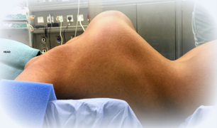 Kyphoscoliosis of the back of someone with kyphoscoliosis EDS
 -
 Severe joint hypermobility in a girl with EDS arthrochalasia type
 -
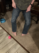 Male, late adolescent, with hypermobile type


Skin
[edit]The weak connective tissue causes abnormal skin. This may present as stretchy or in other types simply be velvet soft. In all types, some increased fragility occurs, but the degree varies depending on the underlying subtype. The skin may tear and bruise easily, and may heal with abnormal atrophic scars;[44] atrophic scars that look like cigarette paper are a sign seen including in those whose skin might appear otherwise normal.[1][15][27] In some subtypes, though not the hypermobile subtype, redundant skin folds occur, especially on the eyelids. Redundant skin folds are areas of excess skin lying in folds.[44][55]
Other skin symptoms include molluscoid pseudotumors,[56] especially on pressure points, petechiae,[57] subcutaneous spheroids,[56] livedo reticularis, and piezogenic papules are less common.[58] In vascular EDS, skin can also be thin and translucent. In dermatosparaxis EDS, the skin is extremely fragile and saggy.[1]
-
 Atrophic scar in a case of EDS
 -
 Translucent skin in vascular EDS
 -
 Individual with EDS displaying skin hyperelasticity
 -
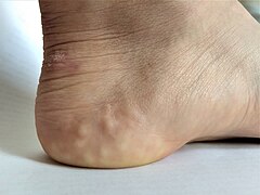 Piezogenic papules on the heel of an individual with hypermobile EDS
 -
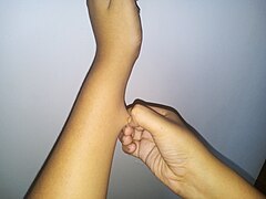 Skin hyperelasticity in the wrist

.png)


Cardiovascular
[edit]- Thoracic outlet syndrome[59]
- Arterial rupture[15]
- Valvular heart disease, such as mitral valve prolapse, creates an increased risk for infective endocarditis during surgery. This may progress to a life-threatening degree.[17] Heart conduction abnormalities have been found in those with hypermobility form of EDS.[60]
- Dilation or rupture (aneurysm) of ascending aorta[61]
- Cardiovascular autonomic dysfunction such as postural orthostatic tachycardia syndrome[62][63][64]
- Raynaud's phenomenon
- Varicose veins[65]
- Heart murmur
- Heart conduction abnormalities
Urogynaecological
[edit]Weakened connective tissues can lead to pelvic organ prolapse in female patients with Ehlers–Danlos syndrome.[66] Patients may also experience voiding difficulties, frequent urinary tract infections, and incontinence due to structural abnormalities.[67] Pelvic girdle pain is also frequently reported.[68]
Menorrhagia, dysmenorrhea, and dyspareunia are common symptoms associated with Ehlers–Danlos syndrome[69] and are often mistaken for endometriosis.[69] Excessive menstrual bleeding can sometimes be attributed to inappropriate platelet aggregation, but faulty collagen leads to weakened capillary walls which increases likelihood of hemorrhage.[70]
In cases of pregnancy, patients with Ehlers–Danlos syndrome are more likely to experience complications during parturition.[70] Post-partum hemorrhage and maternal injury such as sporadic pelvic displacement, hip dislocation, torn and stretched ligaments, and skin tearing can all be linked to altered structure of connective tissues.[71]
Gastrointestinal
[edit]Research suggests a correlation between connective tissue disorders such as Ehlers–Danlos syndrome and both structural and functional problems within the gastrointestinal tract.[72]
Inflammatory problems
[edit]High incidences of coexisting inflammatory disorders suggest a correlation between connective tissue disorders and the development of such aforementioned conditions. Inflammatory bowel diseases such as Crohn's disease, ulcerative colitis[73] and celiac disease[74] are more common in EDS patients when compared to control groups. Of note, patients who are already diagnosed with an inflammatory bowel disorder are not necessarily likely to develop symptoms of a connective tissue disorder, as the two have separate but not totally confounding etiologies.[75] Eosinophilic esophagitis, an inflammatory condition characterized by allergic-type reactions to various foods and chemicals and extensive esophageal remodeling, is eight times more likely in patients with connective tissue disorders when compared to patients without.[76]
Functional problems
[edit]Functionally, small bowel dysmotility, delayed gastric emptying and delayed colonic transit are commonly related to EDS.[77][78] These changes in transit speeds within the gastrointestinal system can cause a host of symptoms, including but not limited to abdominal pain, bloating, nausea, reflux symptoms, vomiting, constipation, and diarrhea.[79]
Some studies also suggest problems with the liver, which is in large part responsible for bilirubin conjugation. Although research in this area is sparse, patients with joint hypermobility were found to have higher rates of indirect hyperbilirubinemia than control groups.[80]
Structural problems
[edit]Structurally, changes within the musculature in the intestine such as increased elastin, can lead to increased frequency of herniation.[72] Laxity of the phreno-esophageal and gastro-hepatic ligaments can lead to hiatal hernia,[81][82] which in turn can lead to commonly reported symptoms such as acid reflux, abdominal pain, early satiety, and bloating. Internal organ prolapses and intestinal intussusceptions occur with greater frequency in patients with weakened connective tissues.[83]
Autonomic problems
[edit]Although neurogastroenterological manifestations in connective tissue disorders are common, their root cause is not yet known.[82] Splanchnic circulation, small fiber neuropathy and altered vascular compliance have all been named as potential contributors to gastrointestinal complaints,[84] particularly for patients who have a known, comorbid autonomic condition.
Neurological
[edit]Chronic headaches are common in patients with Ehlers–Danlos syndrome, whether related to dysautonomia,[85] TMJ, muscle tension, or craniocervical instability. Craniocervical instability is caused by trauma(s) to the head and neck areas such as concussion and whiplash. Ligaments in neck are unable to heal properly, so the neck structure does not have the ability to support the skull, which can then sink into the brain stem, blocking the flow of cerebrospinal fluid, which in turn causes autonomic dysfunction.[86][48]
Arnold–Chiari malformation[87] is also more frequently found in patients with EDS because of the instability at the juncture between skull and spine. This causes herniation of the posterior fossa below the foramen magnum.[88] Increased pressure created by the malformation can lead to a flattened pituitary gland, hormone changes, sudden severe headaches, ataxia, and poor proprioception.[89]
Ophthalmological manifestations include nearsightedness, retinal tearing and retinal detachment, keratoconus, blue sclera, dry eye, Sjogren's syndrome, lens subluxation, angioid streaks, epicanthal folds, strabismus, corneal scarring, brittle cornea syndrome, cataracts, carotid-cavernous sinus fistulas, and macular degeneration.[90]
Otological complications may also occur. Hearing loss is common, both conductive and sensorineural, and is most often bilateral.[91] Otosclerosis and instability of the bones in the inner ear may also contribute to hearing loss[92]
Other manifestations
[edit]- Hiatal hernia[56]
- Gastroesophageal reflux[93]
- Poor gastrointestinal motility[94]
- Dysautonomia[95]
- Gorlin's sign (touch tongue to nose)[96]
- Anal prolapse[56]
- Flat feet
- Tracheobronchomalacia / tracheal collapse
- Collapsed lung (spontaneous pneumothorax)[44]
- Nerve disorders (carpal tunnel syndrome, acroparesthesia, neuropathy, including small fiber neuropathy)[97]
- Insensitivity to local anesthetics[98]
- Dental problems including gum disease and enamel hypoplasia
- Platelet aggregation failure (platelets do not clump together properly)[99]
- Mast cell disorders (including mast cell activation syndrome and mastocytosis)[100]
- Pregnancy complications: increased pain, mild to moderate peripartum bleeding, cervical insufficiency, uterine tearing,[46] or premature rupture of membranes[101]
- Hearing loss may occur in some types[102]
- There is some evidence that EDS may be associated with greater than expected frequencies of neurodevelopmental disorders such as attention deficit hyperactivity disorder (ADHD) and other learning, communication and motor issues, including autism spectrum conditions and Tourette syndrome.[103]
-
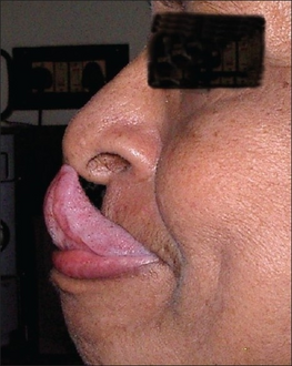 Gorlin's sign in a case of EDS
 -
 Keratoglobus in a case of EDS with brittle cornea syndrome


Because it is often undiagnosed or misdiagnosed in childhood, some instances of EDS have been mischaracterized as child abuse.[104] The pain may also be misdiagnosed as a behavior disorder or Munchausen by proxy.[105]
The pain associated with EDS ranges from mild to debilitating.[106]
Causes
[edit]
Every type of EDS except the hypermobile type (which affects the vast majority of people with EDS) can be positively tied to specific genetic variation[citation needed].
Variations in these genes can cause EDS:[8]
- Collagen primary structure and collagen processing: ADAMTS2, COL1A1, COL1A2, COL3A1, COL5A1, COL5A2
- Collagen folding and collagen cross-linking: PLOD1, FKBP14
- Structure and function of myomatrix: TNXB, COL12A1
- Glycosaminoglycan biosynthesis: B4GALT7, B3GALT6, CHST14, DSE
- Complement pathway: C1R, C1S
- Intracellular processes: SLC39A13, ZNF469, PRDM5
Variations in these genes usually alter the structure, production, or processing of collagen or proteins that interact with collagen. Collagen provides structure and strength to connective tissue. A defect in collagen can weaken connective tissue in the skin, bones, blood vessels, and organs, resulting in the features of the disorder.[1] Inheritance patterns depend on the specific syndrome.
Most forms of EDS are inherited in an autosomal dominant pattern, which means only one of the two copies of the gene in question must be altered to cause a disorder. A few are inherited in an autosomal recessive pattern, which means both copies of the gene must be altered for a person to be affected. It can also be an individual (de novo or "sporadic") variation. Sporadic variations occur without any inheritance.[107]
Diagnosis
[edit]A diagnosis can be made by an evaluation of medical history and clinical observation. The Beighton criteria are widely used to assess the degree of joint hypermobility. DNA and biochemical studies can help identify affected people. Diagnostic tests include collagen gene-variant testing, collagen typing via skin biopsy, echocardiogram, and lysyl hydroxylase or oxidase activity, but these tests are not able to confirm all cases, especially in instances of an unmapped variation, so clinical evaluation remains important. If multiple members of a family are affected, prenatal diagnosis may be possible using a DNA information technique known as a linkage study.[108] Knowledge about EDS among all kinds of practitioners is poor.[109][110] Research is ongoing to identify genetic markers for all types.[111]
Diagnosing hEDS
[edit]Because no single gene has been identified as the sole cause of the most common type of EDS, hypermobile type, obtaining a diagnosis is often difficult[112]. The 2017 diagnositic criteria are as follows:
- Criterion 1: Generalized joint hypermobility, as measured by the Beighton score
- Criterion 2: Minimum two of the following must be met:
- Symptoms that suggest a difference in connective tissue structure
- Unusually soft or velvety skin
- Mild skin hyperextensibility
- Unexplained striae distensae or rubae at the back, groins, thighs, breasts and/or abdomen in adolescents, men or pre-pubertal women without a history of significant gain or loss of body fat or weight
- Bilateral piezogenic papules of the heel
- Recurrent or multiple abdominal hernia(s)
- Atrophic scarring involving at least two sites and without the formation of truly papyraceous and/or hemosideric scars as seen in classical EDS
- Pelvic floor, rectal, and/or uterine prolapse in children, men or nulliparous women without a history of morbid obesity or other known predisposing medical condition
- Dental crowding and high or narrow palate
- Arachnodactyly
- Arm span-to-height ratio ≥1.05
- Mitral valve prolapse (MVP) mild or greater based on strict echocardiographic criteria
- Aortic root dilatation with Z-score >+2
- Positive family history
- Proof that these symptoms interfere with daily life
- Musculoskeletal pain in two or more limbs, recurring daily for at least 3 months
- Chronic, widespread pain for ≥3 months
- Recurrent joint dislocations or frank joint instability, in the absence of trauma
- Symptoms that suggest a difference in connective tissue structure
- Criterion 3: Exclusion of all other possible connective tissue disorders that may be the root cause of symptoms.
In such cases where all criterion are met, a patient can be diagnosed with hEDS according to The International Consortium on Ehlers-Danlos Syndromes and Hypermobility Spectrum Disorders[113]
Differential diagnosis
[edit]Several disorders share some characteristics with EDS. For example, in cutis laxa, the skin is loose, hanging, and wrinkled. In EDS, the skin can be pulled away from the body, but is elastic and returns to normal when let go. In Marfan syndrome, the joints are very mobile and similar cardiovascular complications occur. People with a "marfanoid" appearance are often tall and thin with long arms and legs and "spidery" fingers while EDS phenotypes vary considerably. Certain subtypes of EDS may involve short stature, large eyes, and the appearance of a small mouth and chin, due to a small palate. The palate can have a high arch, causing dental crowding. Blood vessels can sometimes be easily seen through translucent skin, especially on the chest. The genetic connective tissue disorder Loeys–Dietz syndrome also has symptoms that overlap with EDS.[114]
In the past, Menkes disease, a copper metabolism disorder, was thought to be a form of EDS. People are commonly misdiagnosed with fibromyalgia, bleeding disorders, or other disorders that can mimic EDS symptoms. Because of these similar disorders and complications that can arise from an unmonitored case of EDS, a correct diagnosis is important.[115] Pseudoxanthoma elasticum is worth consideration in diagnosis.[116]
Management
[edit]No cure for Ehlers–Danlos syndrome is known, and treatment is supportive. Close monitoring of the cardiovascular system, physiotherapy, occupational therapy, and orthopedic instruments (e.g., wheelchairs, bracing, casting) may be helpful. This can help stabilize the joints and prevent injury. Orthopedic instruments are helpful for the prevention of further joint damage, especially for long distances. People should avoid activities that cause the joint to lock or overextend.[117]
A physician may prescribe casting to stabilize joints. Physicians may refer a person to an orthotist for orthotic treatment (bracing). Physicians may also consult a physical and/or occupational therapist to help strengthen muscles and to teach people how to properly use and preserve their joints.[118][119]
Aquatic therapy promotes muscular development and coordination.[120] With manual therapy, the joint is gently mobilized within the range of motion and/or manipulations.[118][119] If conservative therapy is not helpful, surgical joint repair may be necessary. Medication to decrease pain or manage cardiac, digestive, or other related conditions may be prescribed. To decrease bruising and improve wound healing, some people have responded to vitamin C.[121] Medical care workers often take special precautions because of the sheer number of complications that tend to arise in people with EDS. In vascular EDS, signs of chest or abdominal pain are considered trauma situations.[122]
Cannabinoids and medical marijuana have shown some efficacy in reducing pain levels.[123]
In general, medical intervention is limited to symptomatic therapy. Before pregnancy, people with EDS may be recommended to have genetic counseling and to familiarize themselves with the risks pregnancy poses. Children with EDS should be given information about the disorder so they can understand why they should avoid contact sports and other physically stressful activities. Children should be taught that they should not demonstrate the unusual positions they can maintain due to loose joints, as this may cause early degeneration of the joints. Emotional support along with behavioral and psychological therapy can be useful. Support groups can be immensely helpful for people dealing with major lifestyle changes and poor health. Family members, teachers, and friends should be informed about EDS so they can accept and assist the child.[124]
Pain management
[edit]Successful treatment of chronic pain in EDS requires a multidisciplinary team. The ways to manage pain can be to modify pain management techniques used in the normal population. Pain is classified into several types. One is nociceptive, which is caused by an injury sustained to tissues. Another is neuropathic pain, caused by abnormal signals by the nervous system. In many cases, the pain individuals experience is an unequal mix of the two. Physiotherapy (exercise rehabilitation) can be helpful, especially stabilizing the core and the joints. Stretching exercises must be reduced to slow and gentle stretching to reduce the risks of dislocations or subluxations. Usable methods may include posture reeducation, muscle release, joint mobilization, trunk stabilization, and manual therapy for overworked muscles. Cognitive behavioural therapy is used in many chronic pain patients, especially those who have severe, chronic, life-controlling pain that is unresponsive to treatment. It has not been checked for efficiency in clinical trials. The state of pain management with EDS is considered insufficient.[105]
Medications
[edit]Nonsteroidal anti-inflammatory drugs (NSAIDs) may help if the pain is caused by inflammation. But long-term use of NSAIDs is often a risk factor for gastrointestinal, renal, and blood-related side effects. It can worsen symptoms of mast cell activation syndrome, a disease that may be associated with EDS. Acetaminophen can be used to avoid the bleeding-related side effects of NSAIDs.[105]
Lidocaine can be applied topically after subluxations and painful gums. It can also be injected into painful areas in the case of musculoskeletal pain.[105]
If the pain is neuropathic in origin, tricyclic antidepressants in low doses, anticonvulsants, and selective norepinephrine reuptake inhibitors can be used.[105]
Dislocation and subluxation management
[edit]When a dislocation or subluxation does occur, muscle spasms and stress tend to occur, increasing pain and reducing the chances of the dislocation/subluxation naturally relieving. Methods to support a joint after such an incident include usage of a sling to hold the joint in place and allow it to relax. Orthopedic casts are not advised as there could be pain if unrelaxed muscles are still trying to spasm out against the cast. Other solutions to promote relaxation are heat, gentle massaging, and mental distractions.[125]
Surgery
[edit]The instability of joints, leading to subluxations and joint pain, often requires surgical intervention in people with EDS. Instability of almost all joints can happen, but appears most often in the lower and upper extremities, with the wrist, fingers, shoulder, knee, hip, and ankle being most common.[118]
Common surgical procedures are joint debridement, tendon replacements, capsulorrhaphy, and arthroplasty. After surgery, the degree of stabilization, pain reduction, and people's satisfaction can improve, but surgery does not guarantee an optimal result; affected peoples and surgeons report being dissatisfied with the results. The consensus is that conservative treatment is more effective than surgery,[60] particularly since people have extra risks of surgical complications due to the disease. Three basic surgical problems arise due to EDS – the strength of the tissues is decreased, which makes the tissue less suitable for surgery; the fragility of the blood vessels can cause problems during surgery; and wound healing is often delayed or incomplete.[118] If considering surgical intervention, seeking care from a surgeon with extensive knowledge and experience in treating people with EDS and joint hypermobility issues would be prudent.[126]
Local anesthetics, arterial catheters, and central venous catheters cause a higher risk of bruise formation in people with EDS. Some people with EDS also show a resistance to local anaesthetics.[127] Resistance to lidocaine and bupivacaine is not uncommon, and mepivacaine tends to work better in people with EDS. Special recommendations for anesthesia are given for people with EDS.[citation needed] Detailed recommendations for anesthesia and perioperative care of people with EDS should be used to improve safety.[126]
Surgery in people with EDS requires careful tissue handling and a longer immobilization afterward.[128]
Prognosis
[edit]The outcome for individuals with EDS depends on the specific type of EDS they have. Symptoms vary in severity, even in the same disorder, and the frequency of complications varies. Some people have negligible symptoms, while others are severely restricted in daily life. Extreme joint instability, chronic musculoskeletal pain, degenerative joint disease, frequent injuries, and spinal deformities may limit mobility. Severe spinal deformities may affect breathing. In the case of extreme joint instability, dislocations may result from simple tasks such as rolling over in bed or turning a doorknob. Secondary conditions such as autonomic dysfunction or cardiovascular problems, occurring in any type, can affect prognosis and quality of life. Severe mobility-related disability is seen more often in hEDS than in classical EDS or vascular EDS.[129]
Although all types of EDS are potentially life-threatening,[10][11][12] most people have a normal lifespan. Those with blood vessel fragility, though, have a high risk of fatal complications, including spontaneous arterial rupture, which is the most common cause of sudden death. The median life expectancy in the population with vascular EDS is 48 years.[130]
Complications
[edit]Vascular
[edit]- Pseudoaneurysm[131]
- Vascular lesions (nature is disputed) due to tears in the lining of the arteries or deterioration of congenitally thin and fragile tissue[131]
- Enlarged arteries[131]
Gastrointestinal
[edit]- A 50% risk of colonic perforation exists in vascular Ehlers–Danlos syndrome.[131]
Obstetric
[edit]- Pregnancy increases the likelihood of uterine rupture.[131]
- Maternal mortality is around 12% for the vascular type.[131]
- Uterine hemorrhage can occur during the postpartum recovery.[131]
Epidemiology
[edit]Ehlers–Danlos syndromes are estimated to occur in about one in 5,000 births worldwide. Initially, prevalence estimates ranged from one in 250,000 to 500,000 people, but these were soon found to be low, as medical professionals became more adept at diagnosis. EDS may be far more common than the currently accepted estimate due to the wide range of severities with which the disorder presents.[132]
The prevalence of the disorders differs dramatically. The most common is hypermobile EDS, followed by classical EDS. The others are very rare. For example, fewer than 10 infants and children with dermatosparaxis EDS have been described worldwide.
Some types of EDS are more common in Ashkenazi Jews. For example, the chance of being a carrier for dermatosparaxis EDS is one in 2,000 in the general population but one in 248 among Ashkenazi Jews.[133]
History
[edit]Until 1997, the classification system for EDS included ten specific types and acknowledged that other rarer types existed. At this time, the classification system underwent an overhaul and was reduced to six major types using descriptive titles. Genetic specialists recognize that other types of this condition exist but have only been documented in single families. Except for hypermobility (type 3), the most common type of all ten types, some of the specific variations involved have been identified, and they can be precisely identified by genetic testing; this is valuable due to a great deal of variation in individual cases. Negative genetic test results do not rule out the diagnosis since not all variations have been discovered; therefore, the clinical presentation is crucial.[134]
Forms of EDS in this category may present with soft, mildly stretchable skin, shortened bones, chronic diarrhea, joint hypermobility and dislocation, bladder rupture, or poor wound healing. Inheritance patterns in this group include X-linked recessive, autosomal dominant, and autosomal recessive. Examples of types of related syndromes other than those above reported in the medical literature include:[135]
- 305200: type 5
- 130080: type 8 – unspecified gene, locus 12p13
- 225310: type 10 – unspecified gene, locus 2q34
- 608763: Beasley–Cohen type
- 130070: progeroid form – B4GALT7
- 130090: type unspecified
- 601776: D4ST1-deficient Ehlers–Danlos syndrome (adducted thumb-clubfoot syndrome) CHST14
Society and culture
[edit]EDS may have contributed to the virtuoso violinist Niccolò Paganini's skill, as he was able to play wider fingerings than a typical violinist.[136]
Many sideshow performers have EDS. Several of them were billed as the Elastic Skin Man, the India Rubber Man, and Frog Boy. They included such well-known individuals (in their time) as Felix Wehrle, James Morris, and Avery Childs. Two performers with EDS currently hold world records. Contortionist Daniel Browning Smith has hypermobile EDS and holds the current Guinness World Record for the most flexible man as of 2018, while Gary "Stretch" Turner, sideshow performer in the Circus Of Horrors, has held the current Guinness World Record for the most elastic skin since 1999, for his ability to stretch the skin on his stomach 6.25 inches.[137]
Notable cases
[edit]
- Actress Cherylee Houston has hypermobile EDS. She uses a wheelchair and was the first full-time disabled actress on Coronation Street[138]
- Drag queen and winner of the 11th season of RuPaul's Drag Race Yvie Oddly[139]
- Eric the Actor, a regular caller to The Howard Stern Show[140]
- Actress and activist Jameela Jamil[141]
- Writer and actress Lena Dunham[142]
- Australian singer Sia[143]
- Singer Halsey[144]
- YouTuber and disability rights activist Annie Elainey[145]
- Miss America 2020 Camille Schrier[146]
- Baroque flutist, musicologist and broadcast classical music presenter Hannah French[147]
- Deaf singer/songwriter Mandy Harvey
- YouTuber and disability rights activist Jessica Kellgren-Fozard.[148]
- Singer-songwriter, author and artist Martha Marlow[149]
- Pornographic actor and writer Lorelei Lee[150]
- Reality show contestant Trevor Jones, dead from the vascular form of the disorder[151]
- Scrunchie creator Rommy Hunt Revson, dead from a ruptured aorta[152]
- Japanese voice actress and singer Tomori Kusunoki[153]
Representation in media
[edit]Literature
[edit]The fantasy novel Fourth Wing by Rebecca Yarros presents a main character, Violet Sorrengail, who has an unnamed chronic condition that aligns closely with EDS symptoms. When asked about this connection Rebecca Yarros agrees with the connection but says EDS goes unnamed due to the level of medical knowledge present in her story's world. Yarros has EDS and included it as representation of her condition.[154]
Television
[edit]Grey's Anatomy, a long running TV-series, approached the topic of EDS in their 13th season. In the episode "Falling Slowly", the show's doctors are confronted with confusion when met with diagnosing a patient. Due to complex and contradicting symptoms presented by the patient the show's doctors ultimately give the diagnosis of EDS. This episode was based on conversations held by producers who talked with a patient and doctor who have EDS.[155]
Other species
[edit]Ehlers–Danlos-like syndromes have been shown to be hereditary in Himalayan cats, some domestic shorthair cats,[156] and certain breeds of cattle.[157] It is seen as a sporadic condition in domestic dogs, with higher frequency in English Springers.[158] It has a similar treatment and prognosis. Animals with the condition should not be bred, as the condition can be inherited.[159]
- Animal EDS
-
 EDS in a dog
 -
 Same dog with EDS
 -
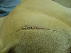 EDS in the same dog showing an atrophic scar


Degenerative suspensory ligament desmitis is a similar condition seen in many breeds of horses.[160] It was originally notated in the Peruvian Paso and thought to be a condition of overwork and older age, but it is being recognized in all age groups and all activity levels. It has been noted in newborn foals.
See also
[edit]References
[edit]- ^ a b c d e f g h i j k l m n o p "Ehlers–Danlos syndrome". MedlinePlus. U.S. National Library of Medicine. Archived from the original on 8 May 2016. Retrieved 9 January 2024.
The 2017 classification describes 13 types of Ehlers–Danlos syndrome.
- ^ "Amplified Musculoskeletal Pain Syndrome (AMPS)". Children's Health.
- ^ a b Anderson BE (2012). The Netter Collection of Medical Illustrations – Integumentary System (2nd ed.). Elsevier Health Sciences. p. 235. ISBN 978-1455726646. Archived from the original on 2017-11-05 – via E-Book.
- ^ a b c d e f g h i j Lawrence EJ (December 2005). "The clinical presentation of Ehlers–Danlos syndrome". Advances in Neonatal Care. 5 (6): 301–314. doi:10.1016/j.adnc.2005.09.006. PMID 16338669. S2CID 7717730.
- ^ a b c d e f g h i j k l m n "Ehlers–Danlos syndromes". Genetic and Rare Diseases Information Center (GARD) – an NCATS Program. U.S. National Institutes of Health. 20 April 2017. Archived from the original on 24 September 2017. Retrieved 23 September 2017.
 This article incorporates text from this source, which is in the public domain.
- ^ a b c Ferri FF (2016). Ferri's Netter Patient Advisor. Elsevier Health Sciences. p. 939. ISBN 9780323393249. Archived from the original on 2017-11-05.
- ^ Dattagupta A, Williamson S, El Nihum LI, Petak S (2022-11-01). "A Case of Spondylodysplastic Ehlers–Danlos Syndrome With Comorbid Hypophosphatasia". AACE Clinical Case Reports. 8 (6): 255–258. doi:10.1016/j.aace.2022.08.005. PMC 9701907. PMID 36447830.
- ^ a b c d e Malfait F, Francomano C, Byers P, Belmont J, Berglund B, Black J, et al. (March 2017). "The 2017 international classification of the Ehlers–Danlos syndromes". American Journal of Medical Genetics. Part C, Seminars in Medical Genetics. 175 (1): 8–26. doi:10.1002/ajmg.c.31552. PMID 28306229. S2CID 4440499.

- ^ a b "Genetics and Inheritance of EDS and HSD". The Ehlers–Danlos Society. 2023.
- ^ a b c Brady AF, Demirdas S, Fournel-Gigleux S, Ghali N, Giunta C, Kapferer-Seebacher I, et al. (March 2017). "The Ehlers–Danlos syndromes, rare types". American Journal of Medical Genetics. Part C, Seminars in Medical Genetics. 175 (1): 70–115. doi:10.1002/ajmg.c.31550. PMID 28306225. S2CID 4439633.
- ^ a b Doolan BJ, Lavallee ME, Hausser I, Schubart JR, Michael Pope F, Seneviratne SL, et al. (23 Jan 2023). "Extracutaneous features and complications of the Ehlers–Danlos syndromes: A systematic review". Frontiers in Medicine. 10: 1053466. doi:10.3389/fmed.2023.1053466. PMC 9899794. PMID 36756177.
- ^ a b Marathe N, Lohkamp LN, Fehlings MG (December 2022). "Spinal manifestations of Ehlers–Danlos syndrome: a scoping review". Journal of Neurosurgery. Spine. 37 (6): 783–793. doi:10.3171/2022.6.SPINE211011. PMID 35986728. S2CID 251694109.
- ^ Tinkle B, Castori M, Berglund B, Cohen H, Grahame R, Kazkaz H, Levy H (March 2017). "Hypermobile Ehlers–Danlos syndrome (a.k.a. Ehlers–Danlos syndrome Type III and Ehlers–Danlos syndrome hypermobility type): Clinical description and natural history". American Journal of Medical Genetics. Part C, Seminars in Medical Genetics. 175 (1): 48–69. doi:10.1002/ajmg.c.31538. PMID 28145611. S2CID 4440630.
- ^ Beighton PH, Grahame R, Bird HA (2011). Hypermobility of Joints. Springer. p. 1. ISBN 9781848820852. Archived from the original on 2017-11-05.
- ^ a b c d Byers PH, Murray ML (November 2012). "Heritable collagen disorders: the paradigm of the Ehlers–Danlos syndrome". The Journal of Investigative Dermatology. 132 (E1): E6-11. doi:10.1038/skinbio.2012.3. PMID 23154631.

- ^ a b c d e f "The Types of EDS". The Ehlers Danlos Society. Retrieved 2019-11-06.
- ^ a b Levy HP (1993). Adam MP, Ardinger HH, Pagon RA, Wallace SE, Bean LJ, Stephens K, Amemiya A (eds.). "Hypermobile Ehlers–Danlos Syndrome". GeneReviews. University of Washington, Seattle. PMID 20301456.
- ^ Carter K. "Hypermobile EDS and Hypermobility Spectrum Disorders". Ehlers–Danlos Support UK.
- ^ "Pregnancy, birth, feeding, and hypermobile Ehlers–Danlos syndrome/hypermobility spectrum disorders". The Ehlers–Danlos Support UK. Retrieved 2019-11-22.
- ^ Cantu L (14 July 2021). "MUSC researchers announce gene mutation discovery associated with EDS". Medical University of South Carolina. Retrieved 2023-02-08.
- ^ "HEDGE Study". ehlers-danlos.com.
- ^ "About the Lab". Norris Lab, Medical University of South Carolina.
- ^ Gensemer C, Burks R, Kautz S, Judge DP, Lavallee M, Norris RA (March 2021). "Hypermobile Ehlers–Danlos syndromes: Complex phenotypes, challenging diagnoses, and poorly understood causes". Developmental Dynamics. 250 (3): 318–344. doi:10.1002/dvdy.220. PMC 7785693. PMID 32629534.
- ^ Narcisi P, Richards AJ, Ferguson SD, Pope FM (September 1994). "A family with Ehlers–Danlos syndrome type III/articular hypermobility syndrome has a glycine 637 to serine substitution in type III collagen". Human Molecular Genetics. 3 (9): 1617–1620. doi:10.1093/hmg/3.9.1617. PMID 7833919.
- ^ Lyons JJ, Yu X, Hughes JD, Le QT, Jamil A, Bai Y, et al. (December 2016). "Elevated basal serum tryptase identifies a multisystem disorder associated with increased TPSAB1 copy number". Nature Genetics. 48 (12): 1564–1569. doi:10.1038/ng.3696. PMC 5397297. PMID 27749843.
- ^ Moore K, Fulmer D, Guo L, Koren N, Glover J, Moore R, et al. (March 2021). "PDGFRα: Expression and Function during Mitral Valve Morphogenesis". Journal of Cardiovascular Development and Disease. 8 (3): 28. doi:10.3390/jcdd8030028. PMC 7999759. PMID 33805717.
- ^ a b c d e Malfait F, Wenstrup R, De Paepe A (July 2018). "Classic Ehlers–Danlos Syndrome". In Adam MP, Ardinger HH, Pagon RA, Wallace SE, Bean LJ, Stephens K, Amemiya A (eds.). GeneReviews. University of Washington, Seattle. PMID 20301422. Retrieved 2019-06-03.
- ^ Kapferer-Seebacher I, Lundberg P, Malfait F, Zschocke J (November 2017). "Periodontal manifestations of Ehlers–Danlos syndromes: A systematic review". Journal of Clinical Periodontology. 44 (11): 1088–1100. doi:10.1111/jcpe.12807. PMID 28836281. S2CID 36252998.
- ^ Castori M (2012). "Ehlers–Danlos syndrome, hypermobility type: an underdiagnosed hereditary connective tissue disorder with mucocutaneous, articular, and systemic manifestations". ISRN Dermatology. 2012: 751768. doi:10.5402/2012/751768. PMC 3512326. PMID 23227356.
- ^ Rakhmanov Y, Maltese PE, Bruson A, Castori M, Beccari T, Dundar M, Bertelli M (2018-09-01). "Genetic testing for vascular Ehlers–Danlos syndrome and other variants with fragility of the middle arteries". The EuroBiotech Journal. 2 (s1): 42–44. doi:10.2478/ebtj-2018-0034. ISSN 2564-615X. S2CID 86589984.
- ^ a b Eagleton MJ (December 2016). "Arterial complications of vascular Ehlers–Danlos syndrome". Journal of Vascular Surgery. 64 (6): 1869–1880. doi:10.1016/j.jvs.2016.06.120. PMID 27687326.
- ^ "Kyphoscoliotic Ehlers–Danlos syndrome". Genetic and Rare Diseases Information Center (GARD) – an NCATS Program. U.S. National Institutes of Health. Retrieved 2019-06-03.
- ^ Klaassens M, Reinstein E, Hilhorst-Hofstee Y, Schrander JJ, Malfait F, Staal H, et al. (August 2012). "Ehlers–Danlos arthrochalasia type (VIIA-B) – expanding the phenotype: from prenatal life through adulthood". Clinical Genetics. 82 (2): 121–130. doi:10.1111/j.1399-0004.2011.01758.x. PMC 4026000. PMID 21801164.
- ^ "Dermatosparaxis Ehlers–Danlos syndrome". Genetic and Rare Diseases Information Center (GARD) – an NCATS Program. U.S. National Institutes of Health. Retrieved 2019-06-03.
- ^ a b "Brittle cornea syndrome". Genetic and Rare Diseases Information Center (GARD) – an NCATS Program. U.S. National Institutes of Health. Retrieved 2019-06-03.
- ^ "OMIM Entry – # 614170 – Brittle Cornea Syndrome 2; BCS2". www.omim.org. Retrieved 2018-06-18.
- ^ "Spondylodysplastic Ehlers–Danlos syndrome". Genetic and Rare Diseases Information Center (GARD) – an NCATS Program. U.S. National Institutes of Health. Archived from the original on 2020-07-08. Retrieved 2019-09-22.
- ^ "Ehlers–Danlos syndrome, musculocontractural type - Conditions – GTR – NCBI". www.ncbi.nlm.nih.gov. Retrieved 2019-09-22.
- ^ Nitahara-Kasahara Y, Mizumoto S, Inoue YU, Saka S, Posadas-Herrera G, Nakamura-Takahashi A, et al. (December 2021). "A new mouse model of Ehlers–Danlos syndrome generated using CRISPR/Cas9-mediated genomic editing". Disease Models & Mechanisms. 14 (12): dmm048963. doi:10.1242/dmm.048963. PMC 8713987. PMID 34850861.
- ^ Guarnieri V, Morlino S, Di Stolfo G, Mastroianno S, Mazza T, Castori M (May 2019). "Cardiac valvular Ehlers–Danlos syndrome is a well-defined condition due to recessive null variants in COL1A2". American Journal of Medical Genetics. Part A. 179 (5): 846–851. doi:10.1002/ajmg.a.61100. PMID 30821104. S2CID 73470267.
- ^ "What are the Ehlers–Danlos Syndromes?". The Ehlers Danlos Society. Retrieved 2021-02-16.
- ^ "Ehlers–Danlos syndrome". Genetic Home Reference. Retrieved 4 April 2018.
- ^ "Ehlers Danlos Syndromes". NORD (National Organization for Rare Disorders). Retrieved 2022-04-19.
- ^ a b c d e "Ehlers–Danlos Syndrome". Mayo Clinic. Archived from the original on 25 June 2012. Retrieved 25 May 2012.
- ^ Wei DH, Terrono AL (October 2015). "Superficialis Sling (Flexor Digitorum Superficialis Tenodesis) for Swan Neck Reconstruction". The Journal of Hand Surgery. 40 (10): 2068–2074. doi:10.1016/j.jhsa.2015.07.018. PMID 26328902.
- ^ a b "Vascular Type-EDS". Ehlers–Danlos Syndrome Network C.A.R.E.S. Inc. Archived from the original on 2012-06-04. Retrieved 2012-05-25.
- ^ Dordoni C, Ciaccio C, Venturini M, Calzavara-Pinton P, Ritelli M, Colombi M (August 2016). "Further delineation of FKBP14-related Ehlers–Danlos syndrome: A patient with early vascular complications and non-progressive kyphoscoliosis, and literature review". American Journal of Medical Genetics. Part A. 170 (8): 2031–2038. doi:10.1002/ajmg.a.37728. hdl:11379/494257. PMID 27149304. S2CID 43512125.
- ^ a b Henderson FC, Austin C, Benzel E, Bolognese P, Ellenbogen R, Francomano CA, et al. (March 2017). "Neurological and spinal manifestations of the Ehlers–Danlos syndromes". American Journal of Medical Genetics. Part C, Seminars in Medical Genetics. 175 (1): 195–211. doi:10.1002/ajmg.c.31549. PMID 28220607. S2CID 4438281.
- ^ Carbone L, Tylavsky FA, Bush AJ, Koo W, Orwoll E, Cheng S (2000). "Bone density in Ehlers–Danlos syndrome". Osteoporosis International. 11 (5): 388–392. doi:10.1007/s001980070104. PMID 10912839. S2CID 441230.
- ^ Nijs J, Van Essche E, De Munck M, Dequeker J (July 2000). "Ultrasonographic, axial, and peripheral measurements in female patients with benign hypermobility syndrome". Calcified Tissue International. 67 (1): 37–40. doi:10.1007/s00223001093. PMID 10908410. S2CID 21083444.
- ^ Gedalia A, Press J, Klein M, Buskila D (July 1993). "Joint hypermobility and fibromyalgia in schoolchildren". Annals of the Rheumatic Diseases. 52 (7): 494–496. doi:10.1136/ard.52.7.494. PMC 1005086. PMID 8346976.
- ^ Dommerholt J (2012-01-27). "CSF Ehlers Danlos Colloquium, Dr Jan Dommerholt". Chiari & Syringomyelia Foundation. Archived from the original on 4 May 2013. Retrieved 10 June 2013.
- ^ Vigorita VJ, Mintz D, Ghelman B (2008). Orthopaedic pathology (2nd ed.). Philadelphia: Lippincott Williams and Wilkins. pp. 5–6. ISBN 978-0781796705.
- ^ "Ehlers–Danlos syndrome - Diagnosis – Approach". BMJ Best Practice. 13 December 2016. Archived from the original on 19 August 2010. Retrieved 18 August 2017.
- ^ "Ehlers Danlos Syndrome – Morphopedics". morphopedics.wikidot.com. Retrieved 2018-06-15.
- ^ a b c d "Classical Type-EDS". Ehlers–Danlos Syndrome Network C.A.R.E.S Inc. Archived from the original on 2012-05-30. Retrieved 2012-05-25.
- ^ Portable Signs and Symptoms. Lippincott Williams & Wilkins. 2007. p. 465. ISBN 9781582556796. Archived from the original on 2017-11-05.
- ^ "Piezogenic papules – DermNet New Zealand". www.dermnetnz.org. Archived from the original on 2016-11-26.
- ^ Ericson WB, Wolman R (March 2017). "Orthopaedic management of the Ehlers–Danlos syndromes". American Journal of Medical Genetics. Part C, Seminars in Medical Genetics. 175 (1): 188–194. doi:10.1002/ajmg.c.31551. eISSN 1552-4876. PMID 28192621. S2CID 4377720.
- ^ a b Camerota F, Castori M, Celletti C, Colotto M, Amato S, Colella A, et al. (July 2014). "Heart rate, conduction and ultrasound abnormalities in adults with joint hypermobility syndrome/Ehlers–Danlos syndrome, hypermobility type". Clinical Rheumatology. 33 (7): 981–987. doi:10.1007/s10067-014-2618-y. PMID 24752348. S2CID 32745869.
- ^ Wenstrup RJ, Hoechstetter LB (2001). "The Ehlers–Danlos Syndromes". In Allanson JE, Cassidy SB (eds.). Management of genetic syndromes. New York: Wiley-Liss. pp. 131–149. ISBN 978-0-471-31286-4.
- ^ Grigoriou E, Boris JR, Dormans JP (February 2015). "Postural orthostatic tachycardia syndrome (POTS): association with Ehlers–Danlos syndrome and orthopaedic considerations". Clinical Orthopaedics and Related Research. 473 (2): 722–728. doi:10.1007/s11999-014-3898-x. eISSN 1528-1132. PMC 4294907. PMID 25156902.
- ^ Hakim A, O'Callaghan C, De Wandele I, Stiles L, Pocinki A, Rowe P (March 2017). "Cardiovascular autonomic dysfunction in Ehlers–Danlos syndrome-Hypermobile type". American Journal of Medical Genetics. Part C, Seminars in Medical Genetics. 175 (1): 168–174. doi:10.1002/ajmg.c.31543. PMID 28160388. S2CID 4439857.
- ^ Ormiston CK, Padilla E, Van DT, Boone C, You S, Roberts AC, Hsiao A, Taub PR (2022-04-09). "May-Thurner syndrome in patients with postural orthostatic tachycardia syndrome and Ehlers-Danlos syndrome: a case series". European Heart Journal: Case Reports. 6 (4): ytac161. doi:10.1093/ehjcr/ytac161. ISSN 2514-2119. PMC 9131024. PMID 35620060.
- ^ Raffetto JD, Khalil RA (April 2008). "Mechanisms of varicose vein formation: valve dysfunction and wall dilation" (PDF). Phlebology. 23 (2): 85–98. doi:10.1258/phleb.2007.007027. PMID 18453484. S2CID 33320026.
- ^ Blagowidow N (December 2021). "Obstetrics and gynecology in Ehlers–Danlos syndrome: A brief review and update". American Journal of Medical Genetics. Part C, Seminars in Medical Genetics. 187 (4): 593–598. doi:10.1002/ajmg.c.31945. PMID 34773390. S2CID 244076387.
- ^ McIntosh LJ, Stanitski DF, Mallett VT, Frahm JD, Richardson DA, Evans MI (1996). "Ehlers–Danlos syndrome: relationship between joint hypermobility, urinary incontinence, and pelvic floor prolapse". Gynecologic and Obstetric Investigation. 41 (2): 135–139. doi:10.1159/000292060. PMID 8838976.
- ^ Kciuk O, Li Q, Huszti E, McDermott CD (February 2023). "Pelvic floor symptoms in natal women with Ehlers–Danlos syndrome: an international survey study". International Urogynecology Journal. 34 (2): 473–483. doi:10.1007/s00192-022-05273-8. PMID 35751670. S2CID 250022004.
- ^ a b Hugon-Rodin J, Lebègue G, Becourt S, Hamonet C, Gompel A (September 2016). "Gynecologic symptoms and the influence on reproductive life in 386 women with hypermobility type ehlers-danlos syndrome: a cohort study". Orphanet Journal of Rare Diseases. 11 (1): 124. doi:10.1186/s13023-016-0511-2. PMC 5020453. PMID 27619482.
- ^ a b Kumskova M, Flora GD, Staber J, Lentz SR, Chauhan AK (July 2023). "Characterization of bleeding symptoms in Ehlers–Danlos syndrome". Journal of Thrombosis and Haemostasis. 21 (7): 1824–1830. doi:10.1016/j.jtha.2023.04.004. PMID 37179130. S2CID 258203231.
- ^ Pearce G, Bell L, Pezaro S, Reinhold E (October 2023). "Childbearing with Hypermobile Ehlers–Danlos Syndrome and Hypermobility Spectrum Disorders: A Large International Survey of Outcomes and Complications". International Journal of Environmental Research and Public Health. 20 (20): 6957. doi:10.3390/ijerph20206957. PMC 10606623. PMID 37887695.
- ^ a b Fikree A, Chelimsky G, Collins H, Kovacic K, Aziz Q (March 2017). "Gastrointestinal involvement in the Ehlers–Danlos syndromes". American Journal of Medical Genetics. Part C, Seminars in Medical Genetics. 175 (1): 181–187. doi:10.1002/ajmg.c.31546. PMID 28186368.
- ^ Adib N, Davies K, Grahame R, Woo P, Murray KJ (June 2005). "Joint hypermobility syndrome in childhood. A not so benign multisystem disorder?". Rheumatology. 44 (6): 744–750. doi:10.1093/rheumatology/keh557. PMID 15728418.
- ^ Danese C, Castori M, Celletti C, Amato S, Lo Russo C, Grammatico P, Camerota F (September 2011). "Screening for celiac disease in the joint hypermobility syndrome/Ehlers–Danlos syndrome hypermobility type". American Journal of Medical Genetics. Part A. 155A (9): 2314–2316. doi:10.1002/ajmg.a.34134. PMID 21815256. S2CID 205314742.
- ^ Laszkowska M, Roy A, Lebwohl B, Green PH, Sundelin HE, Ludvigsson JF (September 2016). "Nationwide population-based cohort study of celiac disease and risk of Ehlers–Danlos syndrome and joint hypermobility syndrome". Digestive and Liver Disease. 48 (9): 1030–1034. doi:10.1016/j.dld.2016.05.019. PMID 27321543.
- ^ Abonia JP, Wen T, Stucke EM, Grotjan T, Griffith MS, Kemme KA, et al. (August 2013). "High prevalence of eosinophilic esophagitis in patients with inherited connective tissue disorders". The Journal of Allergy and Clinical Immunology. 132 (2): 378–386. doi:10.1016/j.jaci.2013.02.030. PMC 3807809. PMID 23608731.
- ^ Zarate N, Farmer AD, Grahame R, Mohammed SD, Knowles CH, Scott SM, Aziz Q (March 2010). "Unexplained gastrointestinal symptoms and joint hypermobility: is connective tissue the missing link?". Neurogastroenterology and Motility. 22 (3): 252–e78. doi:10.1111/j.1365-2982.2009.01421.x. PMID 19840271. S2CID 11516253.
- ^ Menys A, Keszthelyi D, Fitzke H, Fikree A, Atkinson D, Aziz Q, Taylor SA (September 2017). "A magnetic resonance imaging study of gastric motor function in patients with dyspepsia associated with Ehlers–Danlos Syndrome-Hypermobility Type: A feasibility study". Neurogastroenterology and Motility. 29 (9). doi:10.1111/nmo.13090. PMID 28568908. S2CID 776971.
- ^ Zeitoun JD, Lefèvre JH, de Parades V, Séjourné C, Sobhani I, Coffin B, Hamonet C (2013-11-22). Karhausen J (ed.). "Functional digestive symptoms and quality of life in patients with Ehlers–Danlos syndromes: results of a national cohort study on 134 patients". PLOS ONE. 8 (11): e80321. Bibcode:2013PLoSO...880321Z. doi:10.1371/journal.pone.0080321. PMC 3838387. PMID 24278273.
- ^ Çınar M, Çakar M, Öztürk K, Çetindağlı İ, Yılmaz S, Dinç A (March 2017). "Investigation of joint hypermobility in individuals with hyperbilirubinemia". European Journal of Rheumatology. 4 (1): 36–39. doi:10.5152/eurjrheum.2016.16051. PMC 5335885. PMID 28293451.
- ^ Nelson AD, Mouchli MA, Valentin N, Deyle D, Pichurin P, Acosta A, Camilleri M (November 2015). "Ehlers Danlos syndrome and gastrointestinal manifestations: a 20-year experience at Mayo Clinic". Neurogastroenterology and Motility. 27 (11): 1657–1666. doi:10.1111/nmo.12665. PMID 26376608. S2CID 41431319.
- ^ a b Thwaites PA, Gibson PR, Burgell RE (September 2022). "Hypermobile Ehlers–Danlos syndrome and disorders of the gastrointestinal tract: What the gastroenterologist needs to know". Journal of Gastroenterology and Hepatology. 37 (9): 1693–1709. doi:10.1111/jgh.15927. PMC 9544979. PMID 35750466.
- ^ Castori M, Morlino S, Pascolini G, Blundo C, Grammatico P (March 2015). "Gastrointestinal and nutritional issues in joint hypermobility syndrome/Ehlers–Danlos syndrome, hypermobility type". American Journal of Medical Genetics. Part C, Seminars in Medical Genetics. 169C (1): 54–75. doi:10.1002/ajmg.c.31431. PMID 25821092. S2CID 11773627.
- ^ Mehr SE, Barbul A, Shibao CA (August 2018). "Gastrointestinal symptoms in postural tachycardia syndrome: a systematic review". Clinical Autonomic Research. 28 (4): 411–421. doi:10.1007/s10286-018-0519-x. PMC 6314490. PMID 29549458.
- ^ Gazit Y, Nahir A, Grahame R, Jacob G (July 2003). "Dysautonomia in the joint hypermobility syndrome". The American Journal of Medicine. 115 (1): 33–40. doi:10.1016/S0002-9343(03)00235-3. PMID 12867232.
- ^ Henderson F (2015). "Indices of Cranio-vertebral Instability". Funded Research. Chiari & Syringomyelia Foundation. Archived from the original on 2016-09-16.
- ^ Castori M, Voermans NC (October 2014). "Neurological manifestations of Ehlers–Danlos syndrome(s): A review". Iranian Journal of Neurology. 13 (4): 190–208. PMC 4300794. PMID 25632331.
- ^ Dyste GN, Menezes AH, VanGilder JC (1989-08-01). "Symptomatic Chiari malformations: An analysis of presentation, management, and long-term outcome". Journal of Neurosurgery. 71 (2): 159–168. doi:10.3171/jns.1989.71.2.0159. PMID 2746341.
- ^ Langridge B, Phillips E, Choi D (August 2017). "Chiari Malformation Type 1: A Systematic Review of Natural History and Conservative Management". World Neurosurgery. 104: 213–219. doi:10.1016/j.wneu.2017.04.082. ISSN 1878-8750. PMID 28435116.
- ^ William A (18 January 2015). "Ehlers–Danilo's Syndrome – The Role of Collagen in the Eye – Information". The Canadian Ehlers Danlos Association. Archived from the original on 4 April 2016. Retrieved 2019-07-06.
- ^ Weir FW, Hatch JL, Muus JS, Wallace SA, Meyer TA (July 2016). "Audiologic Outcomes in Ehlers-Danlos Syndrome". Otology & Neurotology. 37 (6): 748–752. doi:10.1097/MAO.0000000000001082. ISSN 1531-7129. PMID 27253074. S2CID 5286116.
- ^ Miyajima C, Ishimoto SI, Yamasoba T (January 2007). "Otosclerosis associated with Ehlers-Danlos syndrome: report of a case". Acta Oto-Laryngologica. 127 (sup559): 157–159. doi:10.1080/03655230701600418. ISSN 0001-6489. PMID 18340588. S2CID 2517459.
- ^ Zeitoun JD, Lefèvre JH, de Parades V, Séjourné C, Sobhani I, Coffin B, Hamonet C (November 2013). "Functional digestive symptoms and quality of life in patients with Ehlers–Danlos syndromes: results of a national cohort study on 134 patients". PLOS ONE. 8 (11): e80321. Bibcode:2013PLoSO...880321Z. doi:10.1371/journal.pone.0080321. eISSN 1932-6203. PMC 3838387. PMID 24278273.
- ^ Brockway L (April 2016). "Gastrointestinal manifestations of Ehlers–Danlos syndrome (hypermobility type)". Ehlers–Danlos Support UK. Archived from the original on 2016-11-14.
- ^ "Ehlers–Danlos Syndrome". Underlying Causes of Dysautonomia. Dysautonomia International. 2012. Archived from the original on 2014-12-18.
- ^ Létourneau Y, Pérusse R, Buithieu H (June 2001). "Oral manifestations of Ehlers–Danlos syndrome". Journal. 67 (6): 330–334. PMID 11450296. Archived from the original on 2016-12-15.
- ^ "Ehlers–Danlos syndrome: Definition from". Answers.com. Archived from the original on 2014-03-06. Retrieved 2014-02-27.
- ^ Arendt-Nielsen L. "Patients Suffering from Ehlers Danlos Syndrome Type III Do Not Respond to Local Anesthetics". Archived from the original on 2015-04-05.
- ^ MedlinePlus Encyclopedia: Ehlers–Danlos syndrome
- ^ Seneviratne SL, Maitland A, Afrin L (March 2017). "Mast cell disorders in Ehlers–Danlos syndrome". American Journal of Medical Genetics. Part C, Seminars in Medical Genetics. 175 (1): 226–236. doi:10.1002/ajmg.c.31555. PMID 28261938. S2CID 4438476.
- ^ Lind J, Wallenburg HC (April 2002). "Pregnancy and the Ehlers–Danlos syndrome: a retrospective study in a Dutch population". Acta Obstetricia et Gynecologica Scandinavica. 81 (4): 293–300. doi:10.1034/j.1600-0412.2002.810403.x. hdl:1765/72381. PMID 11952457. S2CID 39943713.
- ^ "Ehlers Danlos Syndromes". NORD (National Organization for Rare Disorders). Retrieved 11 November 2019.
- ^ Baeza-Velasco C (2021). "Neurodevelopmental atypisms in the context of joint hypermobility, hypermobility spectrum disorders, and Ehlers–Danlos syndromes". American Journal of Medical Genetics. Part C, Seminars in Medical Genetics. 187 (4): 491–499. doi:10.1002/ajmg.c.31946. PMID 34741402. S2CID 243801668.
- ^ Santschi DR (April 3, 2008). "Redlands mother stung by untrue suspicions presses for accountability in child abuse inquiries". The Press Enterprise. Archived from the original on February 28, 2009.
- ^ a b c d e Chopra P, Tinkle B, Hamonet C, Brock I, Gompel A, Bulbena A, Francomano C (March 2017). "Pain management in the Ehlers–Danlos syndromes". American Journal of Medical Genetics. Part C, Seminars in Medical Genetics. 175 (1): 212–219. doi:10.1002/ajmg.c.31554. PMID 28186390. S2CID 4433630.
- ^ Chopra P, Tinkle B, Hamonet C, Brock I, Gompel A, Bulbena A, Francomano C (March 2017). "Pain management in the Ehlers–Danlos syndromes". American Journal of Medical Genetics. Part C, Seminars in Medical Genetics. 175 (1): 212–219. doi:10.1002/ajmg.c.31554. PMID 28186390. S2CID 4433630.
- ^ "EDS Types". The Ehlers Danlos Society. Archived from the original on 2017-06-24. Retrieved 2017-05-22.
- ^ Sobey G (January 2015). "Ehlers–Danlos syndrome: how to diagnose and when to perform genetic tests". Archives of Disease in Childhood. 100 (1): 57–61. doi:10.1136/archdischild-2013-304822. PMID 24994860. S2CID 11660992.
- ^ Ross J, Grahame R (January 2011). "Joint hypermobility syndrome". BMJ. 342: c7167. doi:10.1136/bmj.c7167. PMID 21252103. S2CID 1284069.
- ^ Castori M (2012). "Ehlers–danlos syndrome, hypermobility type: an underdiagnosed hereditary connective tissue disorder with mucocutaneous, articular, and systemic manifestations". ISRN Dermatology. 2012: 751768. doi:10.5402/2012/751768. PMC 3512326. PMID 23227356.
- ^ "The Types of EDS". The Ehlers Danlos Society. Retrieved 2018-10-17.
- ^ Forghani I (January 1, 2019). "Updates in Clinical and Genetics Aspects of Hypermobile Ehlers Danlos Syndrome". Balkan Medical Journal. 36 (1) – via National Library of Medicine.
- ^ "The International Consortium on Ehlers-Danlos Syndromes and Hypermobility Spectrum Disorders - The Ehlers Danlos Society". www.ehlers-danlos.com. Retrieved 2024-10-21.
- ^ "Differential Diagnosis". www.loeysdietz.org. Archived from the original on 2017-06-23.
- ^ "Ehlers–Danlos Syndrome". Rarediseases.about.com. 2006-05-25. Archived from the original on 2014-04-12. Retrieved 2014-02-27.
- ^ "Pseudoxanthoma elasticum". Genetics Home Reference. Retrieved 2018-04-17.
- ^ "Physiotherapy and self-management – The Ehlers–Danlos Support UK". www.ehlers-danlos.org. Retrieved 2018-04-17.
- ^ a b c d Rombaut L, Malfait F, De Wandele I, Cools A, Thijs Y, De Paepe A, Calders P (July 2011). "Medication, surgery, and physiotherapy among patients with the hypermobility type of Ehlers–Danlos syndrome". Archives of Physical Medicine and Rehabilitation. 92 (7): 1106–1112. doi:10.1016/j.apmr.2011.01.016. PMID 21636074.
- ^ a b Woerdeman LA, Ritt MJ, Meijer B, Maas M (2000). "Wrist problems in patients with Ehlers–Danlos syndrome". European Journal of Plastic Surgery. 23 (4): 208–210. doi:10.1007/s002380050252. S2CID 29979747.
- ^ Callewaert B, Malfait F, Loeys B, De Paepe A (March 2008). "Ehlers–Danlos syndromes and Marfan syndrome". Best Practice & Research. Clinical Rheumatology. 22 (1): 165–189. doi:10.1016/j.berh.2007.12.005. PMID 18328988.
- ^ Genetics of Ehlers–Danlos Syndrome~treatment at eMedicine
- ^ "Vascular (VEDS) Emergency Information". The Ehlers Danlos Society. Retrieved 2018-04-17.
- ^ "Topics in Pain Management: pain management in patients with hypermobility disorders: frequently missed causes of chronic pain" (PDF). Topics in Pain Management.
- ^ Giroux CM, Corkett JK, Carter LM (2016). "The Academic and Psychosocial Impacts of Ehlers–Danlos Syndrome on Postsecondary Students: An Integrative Review of the Literature" (PDF). Journal of Postsecondary Education and Disability. 29 (4): 414.
- ^ "Dislocation/Subluxation Management". The Ehlers Danlos Society. Retrieved 2022-04-19.
- ^ a b Wiesmann T, Castori M, Malfait F, Wulf H (July 2014). "Recommendations for anesthesia and perioperative management in patients with Ehlers–Danlos syndrome(s)". Orphanet Journal of Rare Diseases. 9: 109. doi:10.1186/s13023-014-0109-5. PMC 4223622. PMID 25053156.
- ^ Parapia LA, Jackson C (April 2008). "Ehlers–Danlos syndrome – a historical review". British Journal of Haematology. 141 (1): 32–35. doi:10.1111/j.1365-2141.2008.06994.x. PMID 18324963. S2CID 7809153.
- ^ Shirley ED, Demaio M, Bodurtha J (September 2012). "Ehlers–danlos syndrome in orthopaedics: etiology, diagnosis, and treatment implications". Sports Health. 4 (5): 394–403. doi:10.1177/1941738112452385. PMC 3435946. PMID 23016112.
- ^ "What are the Ehlers–Danlos Syndromes?". The Ehlers Danlos Society. Retrieved 2019-09-10.
- ^ Pepin M, Schwarze U, Superti-Furga A, Byers PH (March 2000). "Clinical and genetic features of Ehlers–Danlos syndrome type IV, the vascular type". The New England Journal of Medicine. 342 (10): 673–680. CiteSeerX 10.1.1.603.1293. doi:10.1056/NEJM200003093421001. PMID 10706896.
- ^ a b c d e f g Pepin M, Schwarze U, Superti-Furga A, Byers PH (March 2000). "Clinical and genetic features of Ehlers–Danlos syndrome type IV, the vascular type". The New England Journal of Medicine. 342 (10): 673–680. doi:10.1056/NEJM200003093421001. PMID 10706896.
- ^ "Ehlers–Danlos Syndrome: Epidemiology". Medscape.com. Archived from the original on 2013-04-24. Retrieved 2014-02-27.
- ^ "Ehlers–Danlos syndrome type VIIc". geneaware.clinical.bcm.edu. Archived from the original on 2017-08-14. Retrieved 2017-07-24.
- ^ "What is EDS". The Ehlers–Danlos National Foundation. Archived from the original on 2016-04-26. Retrieved 2016-01-06.
- ^ "Ehlers–Danlos syndrome". Online Mendelian Inheritance in Man (OMIM). Retrieved 2019-04-27.
- ^ Yücel D (January 1995). "Was Paganini born with Ehlers–Danlos syndrome phenotype 4 or 3?". Clinical Chemistry. 41 (1): 124–125. doi:10.1093/clinchem/41.1.124. PMID 7813066.
- ^ "Stretchiest skin". Guinness World Records. 29 October 1999. Retrieved 2019-06-22.
- ^ Rollo S (2010-05-22). "Houston hits out at 'preconceived ideas' – Coronation Street News – Soaps". Digital Spy. Archived from the original on 2013-05-09. Retrieved 2014-02-27.
- ^ Oddly Y (April 11, 2019). "Drag Race's Yvie Oddly Opens Up About Living with Ehlers Danos Disease". www.out.com.
- ^ Rosenberg P (30 September 2014). "Eric the Actor: A Eulogy". Rolling Stone. Retrieved 23 July 2019.
- ^ Gillespie C (21 February 2019). "Jameela Jamil Confirms She Has Ehlers–Danlos Syndrome". SELF. Retrieved 9 August 2019.
- ^ Ellis R (November 3, 2019). "Lena Dunham goes on Instagram to reveal she has Ehlers–Danlos syndrome". CNN.
- ^ Doherty J (2019-10-05). "Ehlers–Danlos syndrome: Singer Sia's condition explained". Newsweek. Retrieved 2019-11-11.
- ^ Palowski A (11 May 2022). "What is Ehlers–Danlos syndrome? Halsey reveals diagnosis". Today. Retrieved 19 September 2022.
- ^ Kienapple B (2017-09-01). "Here's What YouTuber Annie Elainey Wants You to Know About Being Disabled". Brit + Co. Retrieved 2019-11-11.
- ^ Bagalawis E (22 July 2019). "Living with little-known disorder Ehlers–Danlos sparked Miss Virginia's love of science". VCU School of Pharmacy News. Retrieved 2020-03-01.
- ^ "Hannah French". Radio 3 Breakfast. BBC. Retrieved 2023-12-25.
- ^ "The Ehlers-Danlos Society Awards". The Ehlers Danlos Society. Retrieved 2024-05-09.
- ^ Skelly S (7 May 2021). "She made us cry with that Qantas song. Now she reveals her own pain". Sydney Morning Herald. Retrieved 2023-02-05.
- ^ "Lorelei Lee's Instagram profile post: "If it seems here on IG like I'm always having another medical procedure that's because it seems that way in real life too. You all are…"". Instagram.com. 2020-11-10. Retrieved 2022-03-03.
- ^ Kubota S (20 October 2021). "Former 'Millionaire Matchmaker' contestant Trevor Jones dies suddenly". Today. Retrieved 2021-10-20.
- ^ Plokhii O (19 September 2022). "WRommy Hunt Revson, who popularized scrunchie, dies at 78". Washington Post. Retrieved 19 September 2022.
- ^ "楠木ともり「エーラス・ダンロス症候群」と公表 『ニジガク』優木せつ菜役は降板" [Tomori Kusunoki announces that she has Ehlers–Danlos syndrome, and quits her role as Setsuna Yuki in 'Nijigaku']. Oricon News (in Japanese). November 2022.
- ^ Straetker R (9 August 2023). "What Condition Does Violet Have in Fourth Wing? Rebecca Yarros Reveals Answer". Health.
- ^ "Ehlers–Danlos syndrome (EDS) on Grey's Anatomy!". EDS Wellness. 14 October 2016.
- ^ Scott DV (October 1974). "Cutaneous asthenia in a cat, resembling Ehlers–Danlos syndrome in man". Veterinary Medicine, Small Animal Clinician. 69 (10): 1256–1258. doi:10.3906/vet-1203-64. PMID 4496767.
- ^ Scott DW (2008). "Congenital and hereditary skin diseases". Color Atlas of Farm Animal Dermatology. Wiley Online Library. p. 61. doi:10.1002/9780470344460. ISBN 9780470344460.
- ^ "English Springer Spaniel - Ehlers–Danlos Syndrome (Cutaneous Asthenia)". Universities Federation for Animal Welfare. Retrieved 28 October 2023.
- ^ Grant D (1 October 2016). "Ehler–Danlos Syndrome (Cutaneous asthenia, dermatosparaxis)". veterinary-practice.com. Retrieved 2019-06-03.
- ^ Halper J (2014). "Connective Tissue Disorders in Domestic Animals". Progress in Heritable Soft Connective Tissue Diseases. Advances in Experimental Medicine and Biology. Vol. 802. pp. 231–240. doi:10.1007/978-94-007-7893-1_14. ISBN 978-94-007-7892-4. PMID 24443030.
External links
[edit]- Cueto I (2022-12-12). "Revenge of the gaslit patients: Now, as scientists, they're tackling Ehlers–Danlos syndromes". STAT. Retrieved 2022-12-13.
- Wan W (December 27, 2021). "A doctor struggled with a rare, incurable syndrome. Now she helps others overcome it". The Washington Post.